 Movie
Movie Controller

[English] 日本語
 Yorodumi
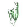
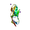
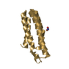
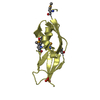
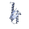
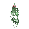
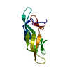
- PDB-4h6h: Crystal Structure of Staphylococcal Complement Inhibitor SCIN-B(4-85) -
+ Open data

- Basic information
| Entry | Database: PDB / ID: 4h6h | ||||||
|---|---|---|---|---|---|---|---|
| Title | Crystal Structure of Staphylococcal Complement Inhibitor SCIN-B(4-85) | ||||||
 Components Components | Fibrinogen-binding protein | ||||||
 Keywords Keywords | IMMUNE SYSTEM / Complement System / Innate Immunity / Staphylococcus aureus Structural Biology | ||||||
| Function / homology |  Function and homology information Function and homology information | ||||||
| Biological species |   Staphylococcus aureus subsp. aureus (bacteria) Staphylococcus aureus subsp. aureus (bacteria) | ||||||
| Method |  X-RAY DIFFRACTION / SYNCHROTRON / MOLECULAR REPLACEMENT / molecular replacement / Resolution: 2.5024 Å X-RAY DIFFRACTION / SYNCHROTRON / MOLECULAR REPLACEMENT / molecular replacement / Resolution: 2.5024 Å | ||||||
 Authors Authors | Garcia, B.L. / Geisbrecht, B.V. | ||||||
 Citation Citation | Journal: J.Biol.Chem. / Year: 2013 Title: A Structurally Dynamic N-terminal Helix Is a Key Functional Determinant in Staphylococcal Complement Inhibitor (SCIN) Proteins. Authors: Garcia, B.L. / Summers, B.J. / Ramyar, K.X. / Tzekou, A. / Lin, Z. / Ricklin, D. / Lambris, J.D. / Laity, J.H. / Geisbrecht, B.V. | ||||||
| History |
|
- Structure visualization
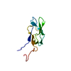
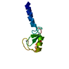
| Structure viewer | Molecule: MolmilJmol/JSmol |
|---|
- Downloads & links
-Download
| PDBx/mmCIF format | 4h6h.cif.gz | 75.6 KB | Display | PDBx/mmCIF format |
|---|---|---|---|---|
| PDB format | pdb4h6h.ent.gz | 57.5 KB | Display | PDB format |
| PDBx/mmJSON format | 4h6h.json.gz | Tree view | PDBx/mmJSON format | |
| Others |  Other downloads Other downloads |
-Validation report
| Arichive directory | https://data.pdbj.org/pub/pdb/validation_reports/h6/4h6hftp://data.pdbj.org/pub/pdb/validation_reports/h6/4h6h | HTTPS FTP |
|---|
-Related structure data
| Related structure data | 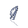 4h6iC 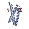 3t49S S: Starting model for refinement C: citing same article ( |
|---|---|
| Similar structure data |
-Links
 PDBj
PDBj
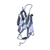
- Assembly
| Deposited unit | 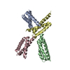
| ||||||||
|---|---|---|---|---|---|---|---|---|---|
| 1 | 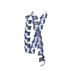
| ||||||||
| 2 | 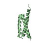
| ||||||||
| 3 | 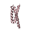
| ||||||||
| 4 | 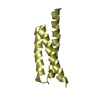
| ||||||||
| Unit cell |
|
-Components
| #1: Protein | Mass: 9980.417 Da / Num. of mol.: 4 Source method: isolated from a genetically manipulated source Source: (gene. exp.) Staphylococcus aureus subsp. aureus (bacteria)Strain: Mu50 / ATCC 700699 / Gene: SAV1159, SAV_1159 / Plasmid: pt7HMT / Production host: #2: Water | ChemComp-HOH / |  Mass: 18.015 Da / Num. of mol.: 63 / Source method: isolated from a natural source / Formula: H2O Mass: 18.015 Da / Num. of mol.: 63 / Source method: isolated from a natural source / Formula: H2O |
|---|
-Experimental details
-Experiment
| Experiment | Method: X-RAY DIFFRACTION / Number of used crystals: 1 |
|---|
- Sample preparation
| Crystal | Density Matthews: 3.77 Å3/Da / Density % sol: 67.35 % |
|---|---|
| Crystal grow | Temperature: 277 K / Method: vapor diffusion, hanging drop / pH: 8.5 Details: 18mg/ml protein sample stored in 10mM Tris (pH 7.4), 50mM NaCl; Crystallization condition: 0.1M Tris (pH 8.5), 8% PEG 8k. Crystals appeared in 1-3 days, VAPOR DIFFUSION, HANGING DROP, temperature 277K |
-Data collection
| Diffraction | Mean temperature: 93 K | |||||||||||||||||||||||||||||||||||||||||||||||||
|---|---|---|---|---|---|---|---|---|---|---|---|---|---|---|---|---|---|---|---|---|---|---|---|---|---|---|---|---|---|---|---|---|---|---|---|---|---|---|---|---|---|---|---|---|---|---|---|---|---|---|
| Diffraction source | Source: SYNCHROTRON / Site: APS  / Beamline: 22-ID / Wavelength: 0.972 Å / Beamline: 22-ID / Wavelength: 0.972 Å | |||||||||||||||||||||||||||||||||||||||||||||||||
| Detector | Type: MARMOSAIC 300 mm CCD / Detector: CCD / Date: Mar 1, 2010 / Details: mirror | |||||||||||||||||||||||||||||||||||||||||||||||||
| Radiation | Monochromator: Si(220) / Protocol: SINGLE WAVELENGTH / Monochromatic (M) / Laue (L): M / Scattering type: x-ray | |||||||||||||||||||||||||||||||||||||||||||||||||
| Radiation wavelength | Wavelength: 0.972 Å / Relative weight: 1 | |||||||||||||||||||||||||||||||||||||||||||||||||
| Reflection | Resolution: 2.5→50 Å / Num. all: 20110 / Num. obs: 17906 / % possible obs: 87.6 % / Observed criterion σ(F): 2 / Observed criterion σ(I): 2 | |||||||||||||||||||||||||||||||||||||||||||||||||
| Reflection shell |
|
-Phasing
| Phasing | Method: molecular replacement |
|---|
- Processing
| Software |
| ||||||||||||||||||||||||||||||||||||||||||||||||||||||||||||||||||||||||||||||||||||||||||||||||||
|---|---|---|---|---|---|---|---|---|---|---|---|---|---|---|---|---|---|---|---|---|---|---|---|---|---|---|---|---|---|---|---|---|---|---|---|---|---|---|---|---|---|---|---|---|---|---|---|---|---|---|---|---|---|---|---|---|---|---|---|---|---|---|---|---|---|---|---|---|---|---|---|---|---|---|---|---|---|---|---|---|---|---|---|---|---|---|---|---|---|---|---|---|---|---|---|---|---|---|---|
| Refinement | Method to determine structure: MOLECULAR REPLACEMENT Starting model: PDB ENTRY 3T49 Resolution: 2.5024→42.262 Å / Occupancy max: 1 / Occupancy min: 1 / FOM work R set: 0.7806 / SU ML: 0.33 / σ(F): 1.97 / Phase error: 28.98 / Stereochemistry target values: ML
| ||||||||||||||||||||||||||||||||||||||||||||||||||||||||||||||||||||||||||||||||||||||||||||||||||
| Solvent computation | Shrinkage radii: 0.9 Å / VDW probe radii: 1.11 Å / Solvent model: FLAT BULK SOLVENT MODEL | ||||||||||||||||||||||||||||||||||||||||||||||||||||||||||||||||||||||||||||||||||||||||||||||||||
| Displacement parameters | Biso max: 128.16 Å2 / Biso mean: 56.2493 Å2 / Biso min: 30 Å2 | ||||||||||||||||||||||||||||||||||||||||||||||||||||||||||||||||||||||||||||||||||||||||||||||||||
| Refinement step | Cycle: LAST / Resolution: 2.5024→42.262 Å
| ||||||||||||||||||||||||||||||||||||||||||||||||||||||||||||||||||||||||||||||||||||||||||||||||||
| Refine LS restraints |
| ||||||||||||||||||||||||||||||||||||||||||||||||||||||||||||||||||||||||||||||||||||||||||||||||||
| LS refinement shell | Refine-ID: X-RAY DIFFRACTION / Total num. of bins used: 13
|
