 Movie
Movie Controller


+ Open data

- Basic information
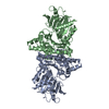
| Entry | Database: PDB / ID: 4h0o | ||||||
|---|---|---|---|---|---|---|---|
| Title | Crystal Structure of Acetate Kinase from Entamoeba histolytica | ||||||
 Components Components | Acetate kinase | ||||||
 Keywords Keywords | TRANSFERASE / pyrophosphate-dependent acetate kinase / ASKHA (acetate and sugar kinase / hsc70 / actin) superfamily / Ribonuclease H-like fold | ||||||
| Function / homology |  Function and homology information Function and homology informationacetate kinase / organic acid metabolic process / acetate kinase activity / acetyl-CoA biosynthetic process / magnesium ion binding / ATP binding Similarity search - Function | ||||||
| Biological species |   Entamoeba histolytica (eukaryote) Entamoeba histolytica (eukaryote) | ||||||
| Method |  X-RAY DIFFRACTION / SYNCHROTRON / MOLECULAR REPLACEMENT / Resolution: 2.4 Å X-RAY DIFFRACTION / SYNCHROTRON / MOLECULAR REPLACEMENT / Resolution: 2.4 Å | ||||||
 Authors Authors | Thaker, T.M. / Tanabe, M. / Iverson, T.M. | ||||||
 Citation Citation | Journal: J.Struct.Biol. / Year: 2013 Title: Crystal structures of acetate kinases from the eukaryotic pathogens Entamoeba histolytica and Cryptococcus neoformans. Authors: Thaker, T.M. / Tanabe, M. / Fowler, M.L. / Preininger, A.M. / Ingram-Smith, C. / Smith, K.S. / Iverson, T.M. | ||||||
| History |
|
- Structure visualization
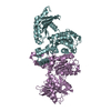
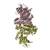
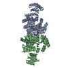
| Structure viewer | Molecule: MolmilJmol/JSmol |
|---|
- Downloads & links
-Download
| PDBx/mmCIF format | 4h0o.cif.gz | 160.8 KB | Display | PDBx/mmCIF format |
|---|---|---|---|---|
| PDB format | pdb4h0o.ent.gz | 127.4 KB | Display | PDB format |
| PDBx/mmJSON format | 4h0o.json.gz | Tree view | PDBx/mmJSON format | |
| Others |  Other downloads Other downloads |
-Validation report
| Summary document | 4h0o_validation.pdf.gz | 434.4 KB | Display | wwPDB validaton report |
|---|---|---|---|---|
| Full document | 4h0o_full_validation.pdf.gz | 440.7 KB | Display | |
| Data in XML | 4h0o_validation.xml.gz | 28.8 KB | Display | |
| Data in CIF | 4h0o_validation.cif.gz | 40 KB | Display | |
| Arichive directory | https://data.pdbj.org/pub/pdb/validation_reports/h0/4h0oftp://data.pdbj.org/pub/pdb/validation_reports/h0/4h0o | HTTPS FTP |
-Related structure data
| Related structure data | 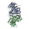 4h0pC 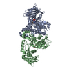 1g99S C: citing same article ( S: Starting model for refinement |
|---|---|
| Similar structure data |


-Links
 PDBj
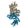
PDBj- Assembly
| Deposited unit | 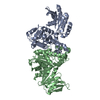
| ||||||||
|---|---|---|---|---|---|---|---|---|---|
| 1 |
| ||||||||
| 2 | 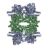
| ||||||||
| Unit cell |
|
-Components
| #1: Protein | Mass: 44703.672 Da / Num. of mol.: 2 Source method: isolated from a genetically manipulated source Source: (gene. exp.) Entamoeba histolytica (eukaryote) / Strain: HM-1:IMSS / Gene: ACKA, EHI_170010 / Plasmid: pQE30 / Production host:  #2: Water | ChemComp-HOH / |  Mass: 18.015 Da / Num. of mol.: 163 / Source method: isolated from a natural source / Formula: H2O Mass: 18.015 Da / Num. of mol.: 163 / Source method: isolated from a natural source / Formula: H2O |
|---|
-Experimental details
-Experiment
| Experiment | Method: X-RAY DIFFRACTION / Number of used crystals: 1 |
|---|
- Sample preparation
| Crystal | Density Matthews: 2.55 Å3/Da / Density % sol: 51.82 % |
|---|---|
| Crystal grow | Temperature: 293 K / Method: vapor diffusion, hanging drop / pH: 6.6 Details: 0.6 M potassium/sodium tartrate, 10 mM iron(III) chloride, 50 mM ADA, pH 6.6, VAPOR DIFFUSION, HANGING DROP, temperature 293K |
-Data collection
| Diffraction | Mean temperature: 100 K |
|---|---|
| Diffraction source | Source: SYNCHROTRON / Site: APS  / Beamline: 21-ID-G / Wavelength: 0.97856 Å / Beamline: 21-ID-G / Wavelength: 0.97856 Å |
| Detector | Type: MARMOSAIC 300 mm CCD / Detector: CCD / Date: Oct 17, 2008 |
| Radiation | Monochromator: Si(111) / Protocol: SINGLE WAVELENGTH / Monochromatic (M) / Laue (L): M / Scattering type: x-ray |
| Radiation wavelength | Wavelength: 0.97856 Å / Relative weight: 1 |
| Reflection | Resolution: 2.4→45.319 Å / Num. all: 44735 / Num. obs: 44735 / % possible obs: 99.3 % / Observed criterion σ(F): 2 / Observed criterion σ(I): 2 / Redundancy: 7.7 % / Rsym value: 0.138 / Net I/σ(I): 15.6 |
| Reflection shell | Resolution: 2.4→2.44 Å / Redundancy: 5.8 % / Mean I/σ(I) obs: 3 / Rsym value: 0.44 / % possible all: 93.7 |
- Processing
| Software |
| ||||||||||||||||||||||||||||||||||||||||||||||||||||||||||||||||||||||||||||||||||||
|---|---|---|---|---|---|---|---|---|---|---|---|---|---|---|---|---|---|---|---|---|---|---|---|---|---|---|---|---|---|---|---|---|---|---|---|---|---|---|---|---|---|---|---|---|---|---|---|---|---|---|---|---|---|---|---|---|---|---|---|---|---|---|---|---|---|---|---|---|---|---|---|---|---|---|---|---|---|---|---|---|---|---|---|---|---|
| Refinement | Method to determine structure: MOLECULAR REPLACEMENT Starting model: PDB ENTRY 1G99 Resolution: 2.4→45.319 Å / SU ML: 0.16 / σ(F): 1.34 / Phase error: 21.56 / Stereochemistry target values: ML
| ||||||||||||||||||||||||||||||||||||||||||||||||||||||||||||||||||||||||||||||||||||
| Solvent computation | Shrinkage radii: 0.9 Å / VDW probe radii: 1.11 Å / Solvent model: FLAT BULK SOLVENT MODEL | ||||||||||||||||||||||||||||||||||||||||||||||||||||||||||||||||||||||||||||||||||||
| Refinement step | Cycle: LAST / Resolution: 2.4→45.319 Å
| ||||||||||||||||||||||||||||||||||||||||||||||||||||||||||||||||||||||||||||||||||||
| Refine LS restraints |
| ||||||||||||||||||||||||||||||||||||||||||||||||||||||||||||||||||||||||||||||||||||
| LS refinement shell |
|
