 Movie
Movie Controller

[English] 日本語
 Yorodumi
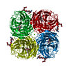
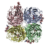
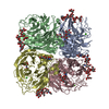
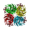
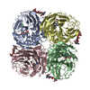
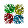
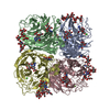
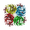
- PDB-4gzo: N2 neuraminidase of A/Tanzania/205/2010 H3N2 in complex with hepes -
+ Open data

- Basic information
| Entry | Database: PDB / ID: 4gzo | |||||||||
|---|---|---|---|---|---|---|---|---|---|---|
| Title | N2 neuraminidase of A/Tanzania/205/2010 H3N2 in complex with hepes | |||||||||
 Components Components | neuraminidase | |||||||||
 Keywords Keywords | VIRAL PROTEIN / beta-propella / infleunza virus / neuraminidase / hemagglutinin / hemadsorption / viral infection / sialic acid receptor | |||||||||
| Function / homology |  Function and homology information Function and homology informationexo-alpha-sialidase / exo-alpha-sialidase activity / viral budding from plasma membrane / carbohydrate metabolic process / host cell plasma membrane / virion membrane / membrane / metal ion binding Similarity search - Function | |||||||||
| Biological species |   Influenza A virus Influenza A virus | |||||||||
| Method |  X-RAY DIFFRACTION / SYNCHROTRON / MOLECULAR REPLACEMENT / Resolution: 2.6 Å X-RAY DIFFRACTION / SYNCHROTRON / MOLECULAR REPLACEMENT / Resolution: 2.6 Å | |||||||||
 Authors Authors | Zhu, X. / Wilson, I.A. | |||||||||
 Citation Citation | Journal: J.Virol. / Year: 2012 Title: Influenza virus neuraminidases with reduced enzymatic activity that avidly bind sialic Acid receptors. Authors: Zhu, X. / McBride, R. / Nycholat, C.M. / Yu, W. / Paulson, J.C. / Wilson, I.A. | |||||||||
| History |
|
- Structure visualization
| Structure viewer | Molecule: MolmilJmol/JSmol |
|---|
- Downloads & links
-Download
| PDBx/mmCIF format | 4gzo.cif.gz | 93.7 KB | Display | PDBx/mmCIF format |
|---|---|---|---|---|
| PDB format | pdb4gzo.ent.gz | 68.9 KB | Display | PDB format |
| PDBx/mmJSON format | 4gzo.json.gz | Tree view | PDBx/mmJSON format | |
| Others |  Other downloads Other downloads |
-Validation report
| Arichive directory | https://data.pdbj.org/pub/pdb/validation_reports/gz/4gzoftp://data.pdbj.org/pub/pdb/validation_reports/gz/4gzo | HTTPS FTP |
|---|
-Related structure data
| Related structure data |  4gzpC  4gzqC  4gzsC 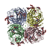 4gztC  4gzwC 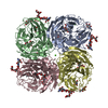 4gzxC 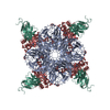 2aepS C: citing same article ( S: Starting model for refinement |
|---|---|
| Similar structure data |


-Links
 PDBj
PDBj


- Assembly
| Deposited unit | 
| ||||||||
|---|---|---|---|---|---|---|---|---|---|
| 1 | 
| ||||||||
| Unit cell |
|
-Components
-Protein , 1 types, 1 molecules A
| #1: Protein | Mass: 43349.352 Da / Num. of mol.: 1 Source method: isolated from a genetically manipulated source Source: (gene. exp.) Influenza A virus / Strain: A/Tanzania/205/2010(H3N2) / Plasmid: PFASTBACHT-A / Production host:  Trichoplusia Ni (cabbage looper) / References: UniProt: K7N5N3*PLUS Trichoplusia Ni (cabbage looper) / References: UniProt: K7N5N3*PLUS |
|---|
-Sugars , 2 types, 3 molecules 
| #2: Polysaccharide | alpha-D-mannopyranose-(1-3)-[alpha-D-mannopyranose-(1-6)]beta-D-mannopyranose-(1-4)-2-acetamido-2- ...alpha-D-mannopyranose-(1-3)-[alpha-D-mannopyranose-(1-6)]beta-D-mannopyranose-(1-4)-2-acetamido-2-deoxy-beta-D-glucopyranose-(1-4)-2-acetamido-2-deoxy-beta-D-glucopyranose Source method: isolated from a genetically manipulated source |
|---|---|
| #4: Sugar |  Type: D-saccharide, beta linking / Mass: 221.208 Da / Num. of mol.: 2 Type: D-saccharide, beta linking / Mass: 221.208 Da / Num. of mol.: 2Source method: isolated from a genetically manipulated source Formula: C8H15NO6 |
-Non-polymers , 3 types, 54 molecules 


| #3: Chemical | ChemComp-EPE /  Mass: 238.305 Da / Num. of mol.: 1 / Source method: obtained synthetically / Formula: C8H18N2O4S / Comment: pH buffer*YM Mass: 238.305 Da / Num. of mol.: 1 / Source method: obtained synthetically / Formula: C8H18N2O4S / Comment: pH buffer*YM | ||
|---|---|---|---|
| #5: Chemical |  Mass: 40.078 Da / Num. of mol.: 2 / Source method: obtained synthetically / Formula: Ca Mass: 40.078 Da / Num. of mol.: 2 / Source method: obtained synthetically / Formula: Ca#6: Water | ChemComp-HOH / | Mass: 18.015 Da / Num. of mol.: 51 / Source method: isolated from a natural source / Formula: H2O |
-Details
| Has protein modification | Y |
|---|---|
| Sequence details | AUTHORS STATE THAT THE GISAID ACCESSION NUMBER IS EPI342198 FOR THIS SEQUENCE. |
-Experimental details
-Experiment
| Experiment | Method: X-RAY DIFFRACTION / Number of used crystals: 1 |
|---|
- Sample preparation
| Crystal | Density Matthews: 2.72 Å3/Da / Density % sol: 54.73 % |
|---|---|
| Crystal grow | Temperature: 295 K / Method: vapor diffusion / pH: 7.5 Details: 0.1 M Hepes, 17-25% PEG 8000, pH 7.5, VAPOR DIFFUSION, temperature 295K |
-Data collection
| Diffraction | Mean temperature: 100 K |
|---|---|
| Diffraction source | Source: SYNCHROTRON / Site: SSRL  / Beamline: BL12-2 / Wavelength: 0.9795 Å / Beamline: BL12-2 / Wavelength: 0.9795 Å |
| Detector | Type: PSI PILATUS 6M / Detector: PIXEL / Date: May 13, 2011 |
| Radiation | Monochromator: liquid nitrogen-cooled double crystal / Protocol: SINGLE WAVELENGTH / Monochromatic (M) / Laue (L): M / Scattering type: x-ray |
| Radiation wavelength | Wavelength: 0.9795 Å / Relative weight: 1 |
| Reflection | Resolution: 2.6→50 Å / Num. obs: 13907 / % possible obs: 90.3 % / Observed criterion σ(I): -3 / Redundancy: 9 % / Biso Wilson estimate: 44.7 Å2 / Rmerge(I) obs: 0.128 |
| Reflection shell | Resolution: 2.6→2.69 Å / Redundancy: 6.6 % / Rmerge(I) obs: 0.34 / % possible all: 64.3 |
- Processing
| Software |
| |||||||||||||||||||||||||||||||||||||||||||||
|---|---|---|---|---|---|---|---|---|---|---|---|---|---|---|---|---|---|---|---|---|---|---|---|---|---|---|---|---|---|---|---|---|---|---|---|---|---|---|---|---|---|---|---|---|---|---|
| Refinement | Method to determine structure: MOLECULAR REPLACEMENT Starting model: PDB entry 2AEP Resolution: 2.6→50 Å / Cor.coef. Fo:Fc: 0.932 / Cor.coef. Fo:Fc free: 0.895 / Cross valid method: THROUGHOUT / ESU R: 2.756 / ESU R Free: 0.367 / Stereochemistry target values: MAXIMUM LIKELIHOOD
| |||||||||||||||||||||||||||||||||||||||||||||
| Solvent computation | Ion probe radii: 0.8 Å / Shrinkage radii: 0.8 Å / VDW probe radii: 1.2 Å / Solvent model: MASK | |||||||||||||||||||||||||||||||||||||||||||||
| Displacement parameters | Biso mean: 46.315 Å2
| |||||||||||||||||||||||||||||||||||||||||||||
| Refinement step | Cycle: LAST / Resolution: 2.6→50 Å
| |||||||||||||||||||||||||||||||||||||||||||||
| Refine LS restraints |
| |||||||||||||||||||||||||||||||||||||||||||||
| LS refinement shell | Resolution: 2.6→2.667 Å / Total num. of bins used: 20
|
