 Movie
Movie Controller

[English] 日本語
 Yorodumi
- PDB-2qwj: THE X-RAY STRUCTURE OF A COMPLEX OF 5-N-ACETYL-4-AMINO-6-DIETHYLC... -
+ Open data

- Basic information
| Entry | Database: PDB / ID: 2qwj | ||||||||||||
|---|---|---|---|---|---|---|---|---|---|---|---|---|---|
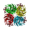
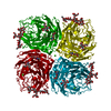
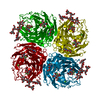
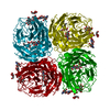
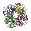
| Title | THE X-RAY STRUCTURE OF A COMPLEX OF 5-N-ACETYL-4-AMINO-6-DIETHYLCARBOXAMIDE-4,5-DIHYDRO-2H-PYRAN-2-CARBOXYLIC ACID AND A DRUG RESISTANT VARIANT R292K OF TERN N9 INFLUENZA VIRUS NEURAMINIDASE | ||||||||||||
 Components Components | NEURAMINIDASE | ||||||||||||
 Keywords Keywords | HYDROLASE / NEURAMINIDASE / INFLUENZA PROTEIN / DRUG RESISTANT VARIANT / SIALIC ACID ANALOGUES / DANA ANOLOGUES / GLYCOSYLATED PROTEIN / CARBOXAMIDE DERIVATIVES | ||||||||||||
| Function / homology |  Function and homology information Function and homology informationexo-alpha-sialidase / exo-alpha-sialidase activity / viral budding from plasma membrane / carbohydrate metabolic process / host cell plasma membrane / virion membrane / membrane / metal ion binding Similarity search - Function | ||||||||||||
| Biological species |   Influenza A virus Influenza A virus | ||||||||||||
| Method |  X-RAY DIFFRACTION / MOLECULAR REPLACEMENT / Resolution: 2 Å X-RAY DIFFRACTION / MOLECULAR REPLACEMENT / Resolution: 2 Å | ||||||||||||
 Authors Authors | Varghese, J.N. | ||||||||||||
 Citation Citation | Journal: Structure / Year: 1998 Title: Drug design against a shifting target: a structural basis for resistance to inhibitors in a variant of influenza virus neuraminidase. Authors: Varghese, J.N. / Smith, P.W. / Sollis, S.L. / Blick, T.J. / Sahasrabudhe, A. / McKimm-Breschkin, J.L. / Colman, P.M. #1: Journal: J.Virol. / Year: 1998Title: Mutations in a Conserved Residue in the Influenza Virus Neuraminidase Active Site Decreases Sensitivity to Neu5Ac2En-Derived Inhibitors Authors: Mckimm-Breschkin, J.L. / Sahasrabudhe, A. / Blick, T.J. / Mcdonald, M. / Colman, P.M. / Hart, G.J. / Bethell, R.C. / Varghese, J.N. | ||||||||||||
| History |
|
- Structure visualization
| Structure viewer | Molecule: MolmilJmol/JSmol |
|---|
- Downloads & links
-Download
| PDBx/mmCIF format | 2qwj.cif.gz | 105.4 KB | Display | PDBx/mmCIF format |
|---|---|---|---|---|
| PDB format | pdb2qwj.ent.gz | 78.1 KB | Display | PDB format |
| PDBx/mmJSON format | 2qwj.json.gz | Tree view | PDBx/mmJSON format | |
| Others |  Other downloads Other downloads |
-Validation report
| Arichive directory | https://data.pdbj.org/pub/pdb/validation_reports/qw/2qwjftp://data.pdbj.org/pub/pdb/validation_reports/qw/2qwj | HTTPS FTP |
|---|
-Related structure data
| Related structure data |  2qwaC  2qwbC  2qwcC  2qwdC  2qweC  2qwfC  2qwgC  2qwhC 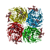 2qwiC  2qwkC C: citing same article ( |
|---|---|
| Similar structure data |


-Links
 PDBj
PDBj

- Assembly
| Deposited unit | 
| |||||||||
|---|---|---|---|---|---|---|---|---|---|---|
| 1 | 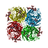
| |||||||||
| Unit cell |
| |||||||||
| Components on special symmetry positions |
|
-Components
-Protein , 1 types, 1 molecules A
| #1: Protein | Mass: 43723.770 Da / Num. of mol.: 1 / Fragment: RESIDUES 82 - 468 Source method: isolated from a genetically manipulated source Details: INTEGRAL MEMBRANE PROTEIN, MEMBRANE BOUND STALK CLEAVED BY PRONASE, RELEASING FULLY ACTIVE HEAD WITH RESIDUES 82 - 468 (TOKYO/3/67 NUMBERING) Source: (gene. exp.) Influenza A virus / Genus: Influenzavirus A / Strain: A/TERN/AUSTRALIA/G70C/75 / Production host:  |
|---|
-Sugars , 4 types, 4 molecules 
| #2: Polysaccharide | alpha-D-mannopyranose-(1-2)-alpha-D-mannopyranose-(1-2)-alpha-D-mannopyranose-(1-3)-[alpha-D- ...alpha-D-mannopyranose-(1-2)-alpha-D-mannopyranose-(1-2)-alpha-D-mannopyranose-(1-3)-[alpha-D-mannopyranose-(1-6)]beta-D-mannopyranose-(1-4)-2-acetamido-2-deoxy-beta-D-glucopyranose-(1-4)-2-acetamido-2-deoxy-beta-D-glucopyranose Source method: isolated from a genetically manipulated source |
|---|---|
| #3: Polysaccharide | 2-acetamido-2-deoxy-beta-D-glucopyranose-(1-4)-2-acetamido-2-deoxy-beta-D-glucopyranose Source method: isolated from a genetically manipulated source |
| #4: Sugar | ChemComp-NAG /  Type: D-saccharide, beta linking / Mass: 221.208 Da / Num. of mol.: 1 Type: D-saccharide, beta linking / Mass: 221.208 Da / Num. of mol.: 1Source method: isolated from a genetically manipulated source Formula: C8H15NO6 |
| #6: Sugar | ChemComp-G28 /  Type: L-saccharide / Mass: 299.323 Da / Num. of mol.: 1 Type: L-saccharide / Mass: 299.323 Da / Num. of mol.: 1Source method: isolated from a genetically manipulated source Formula: C13H21N3O5 |
-Non-polymers , 2 types, 343 molecules 

| #5: Chemical |  Mass: 40.078 Da / Num. of mol.: 2 / Source method: obtained synthetically / Formula: Ca Mass: 40.078 Da / Num. of mol.: 2 / Source method: obtained synthetically / Formula: Ca#7: Water | ChemComp-HOH / | Mass: 18.015 Da / Num. of mol.: 341 / Source method: isolated from a natural source / Formula: H2O |
|---|
-Details
| Has protein modification | Y |
|---|
-Experimental details
-Experiment
| Experiment | Method: X-RAY DIFFRACTION / Number of used crystals: 1 |
|---|
- Sample preparation
| Crystal | Density Matthews: 2.82 Å3/Da / Density % sol: 54 % Description: THIS EXPERIMENT WAS CARRIED OUT TO DETERMINE THE STRUCTURE OF COMPLEX OF 5-N-ACETYL-4-AMINO-6- DIETHYLCARBOXAMIDE-4,5-DIHYDRO-2H-PYRAN-2-CARBOXYLIC ACID AND WILD-TYPE TERN N9 NEURAMINIDASE. | ||||||||||||||||||||
|---|---|---|---|---|---|---|---|---|---|---|---|---|---|---|---|---|---|---|---|---|---|
| Crystal grow | pH: 5.9 / Details: 1.9M PHOSPHATE (PH 5.9) | ||||||||||||||||||||
| Crystal | *PLUS | ||||||||||||||||||||
| Crystal grow | *PLUS pH: 6.6 / Method: vapor diffusion / Details: Laver, W.G., (1984) Virology, 137, 314. | ||||||||||||||||||||
| Components of the solutions | *PLUS
|
-Data collection
| Diffraction | Mean temperature: 107 K |
|---|---|
| Diffraction source | Source: ROTATING ANODE / Type: MACSCIENCE M18X / Wavelength: 1.5418 |
| Detector | Type: RIGAKU RAXIS II / Detector: IMAGE PLATE / Date: Jul 7, 1997 / Details: YALE MIRRORS |
| Radiation | Monochromatic (M) / Laue (L): M / Scattering type: x-ray |
| Radiation wavelength | Wavelength: 1.5418 Å / Relative weight: 1 |
| Reflection | Resolution: 2→100 Å / Num. obs: 32767 / % possible obs: 84 % / Observed criterion σ(I): 1 / Redundancy: 3.2 % / Rmerge(I) obs: 0.125 / Rsym value: 0.121 |
| Reflection shell | Resolution: 2.01→2.1 Å / % possible all: 65.4 |
| Reflection | *PLUS Num. measured all: 106146 |
- Processing
| Software |
| ||||||||||||||||||||||||||||||||||||||||||||||||||||||||||||
|---|---|---|---|---|---|---|---|---|---|---|---|---|---|---|---|---|---|---|---|---|---|---|---|---|---|---|---|---|---|---|---|---|---|---|---|---|---|---|---|---|---|---|---|---|---|---|---|---|---|---|---|---|---|---|---|---|---|---|---|---|---|
| Refinement | Method to determine structure: MOLECULAR REPLACEMENT Starting model: TERN N9 NEURAMINIDASE Resolution: 2→6 Å / σ(F): 1 / Details: 2 METAL IONS
| ||||||||||||||||||||||||||||||||||||||||||||||||||||||||||||
| Refine analyze | Luzzati coordinate error obs: 0.01 Å / Luzzati d res low obs: 6 Å | ||||||||||||||||||||||||||||||||||||||||||||||||||||||||||||
| Refinement step | Cycle: LAST / Resolution: 2→6 Å
| ||||||||||||||||||||||||||||||||||||||||||||||||||||||||||||
| Refine LS restraints |
| ||||||||||||||||||||||||||||||||||||||||||||||||||||||||||||
| LS refinement shell | Resolution: 2→2.03 Å / Total num. of bins used: 20
| ||||||||||||||||||||||||||||||||||||||||||||||||||||||||||||
| Xplor file |
|
