- PDB-4gyt: Crystal structure of lpg0076 protein from Legionella pneumophila -
+
Open data
ID or keywords:
Loading...
-
Basic information
Entry
Database: PDB / ID: 4gyt
Title
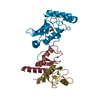
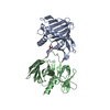
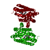
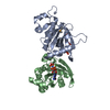
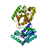
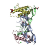
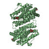
Crystal structure of lpg0076 protein from Legionella pneumophila
Components
uncharacterized protein
Keywords
Structural Genomics / Unknown Function / PSI-Biology / Midwest Center for Structural Genomics / MCSG / predicted Legionella effector
Function / homology
YgfB uncharacterised protein family PF03695 / YgfB-like / YgfB-like superfamily / Uncharacterised protein family (UPF0149) / Four Helix Bundle (Hemerythrin (Met), subunit A) / Up-down Bundle / Mainly Alpha / cytosol / YecA family protein
Monochromator: double crystal / Protocol: SINGLE WAVELENGTH / Monochromatic (M) / Laue (L): M / Scattering type: x-ray
Radiation wavelength
Wavelength: 0.97929 Å / Relative weight: 1
Reflection
Resolution: 2.047→30 Å / Num. all: 29913 / Num. obs: 28918 / % possible obs: 96.7 % / Observed criterion σ(I): -3 / Redundancy: 8.8 % / Biso Wilson estimate: 38.5 Å2 / Rmerge(I) obs: 0.075 / Net I/σ(I): 27.4
Reflection shell
Resolution: 2.05→2.09 Å / Redundancy: 8.8 % / Rmerge(I) obs: 0.752 / Mean I/σ(I) obs: 3.5 / Num. unique all: 1417 / % possible all: 97.3
-
Processing
Software
Name
Version
Classification
SBC-Collect
datacollection
SHELX
modelbuilding
MLPHARE
phasing
DM
modelbuilding
BUCCANEER
modelbuilding
ARP/wARP
modelbuilding
Coot
modelbuilding
PHENIX
(phenix.refine: dev_1096)
refinement
HKL-3000
datareduction
HKL-3000
datascaling
SHELX
phasing
DM
phasing
BUCCANEER
phasing
Refinement
Method to determine structure: SAD / Resolution: 2.047→28.789 Å / SU ML: 0.16 / Isotropic thermal model: isotropic / Cross valid method: THROUGHOUT / σ(F): 0 / Phase error: 21.48 / Stereochemistry target values: ML Details: HYDROGEN ATOMS HAVE BEEN ADDED AT THE RIDING POSITIONS; THE PROTEIN WAS SUBJECTED TO IN SITU PROTEOLYSIS, THEREFORE THE EXACT LENGTH OF THE CRYSTALLIZED POLYPEPTIDE COULD NOT BE DETERMINED
Rfactor
Num. reflection
% reflection
Selection details
Rfree
0.2129
1454
5.04 %
random
Rwork
0.1692
-
-
-
all
0.173
28862
-
-
obs
0.1713
28862
96.44 %
-
Solvent computation
Shrinkage radii: 0.9 Å / VDW probe radii: 1.11 Å / Solvent model: FLAT BULK SOLVENT MODEL
Refinement step
Cycle: LAST / Resolution: 2.047→28.789 Å
Protein
Nucleic acid
Ligand
Solvent
Total
Num. atoms
2798
0
2
122
2922
Refine LS restraints
Refine-ID
Type
Dev ideal
Number
X-RAY DIFFRACTION
f_bond_d
0.012
2918
X-RAY DIFFRACTION
f_angle_d
1.203
3942
X-RAY DIFFRACTION
f_dihedral_angle_d
17.342
1082
X-RAY DIFFRACTION
f_chiral_restr
0.069
426
X-RAY DIFFRACTION
f_plane_restr
0.005
530
LS refinement shell
Resolution (Å)
Rfactor Rfree
Num. reflection Rfree
Rfactor Rwork
Num. reflection Rwork
Refine-ID
% reflection obs (%)
2.0472-2.1203
0.2548
149
0.2091
2666
X-RAY DIFFRACTION
96
2.1203-2.2052
0.2412
148
0.2013
2741
X-RAY DIFFRACTION
98
2.2052-2.3055
0.2357
141
0.1875
2736
X-RAY DIFFRACTION
98
2.3055-2.427
0.234
156
0.1731
2732
X-RAY DIFFRACTION
98
2.427-2.579
0.2542
149
0.1725
2739
X-RAY DIFFRACTION
97
2.579-2.7779
0.2229
134
0.1913
2752
X-RAY DIFFRACTION
97
2.7779-3.0572
0.2717
151
0.2019
2726
X-RAY DIFFRACTION
96
3.0572-3.4989
0.2461
132
0.1782
2733
X-RAY DIFFRACTION
96
3.4989-4.4058
0.1677
157
0.1461
2744
X-RAY DIFFRACTION
95
4.4058-28.7913
0.191
137
0.1564
2839
X-RAY DIFFRACTION
93
Refinement TLS params.
Method: refined / Refine-ID: X-RAY DIFFRACTION
ID
L11 (°2)
L12 (°2)
L13 (°2)
L22 (°2)
L23 (°2)
L33 (°2)
S11 (Å °)
S12 (Å °)
S13 (Å °)
S21 (Å °)
S22 (Å °)
S23 (Å °)
S31 (Å °)
S32 (Å °)
S33 (Å °)
T11 (Å2)
T12 (Å2)
T13 (Å2)
T22 (Å2)
T23 (Å2)
T33 (Å2)
Origin x (Å)
Origin y (Å)
Origin z (Å)
1
3.6434
-0.4273
0.4509
3.4026
-0.4846
3.1631
0.1267
-0.1192
0.1346
-0.0557
-0.2343
-0.3324
-0.2131
0.3723
-0.0131
0.237
-0.0487
-0.0225
0.2503
-0.0085
0.2094
17.0075
29.2838
18.4421
2
0.297
-0.1945
0.1779
0.7637
0.4001
0.5432
-0.1435
-0.6125
-0.8297
0.8875
0.3447
-0.591
0.3355
0.4719
0.0498
0.3025
0.1097
-0.056
0.5398
0.072
0.6538
24.6148
17.4755
22.9323
3
1.4434
0.481
-0.2001
0.6326
-0.445
1.3499
-0.0314
-0.3406
0.2243
0.2615
-0.0443
-0.1723
-0.4463
0.0962
-0.0001
0.3021
-0.0369
-0.0665
0.3011
-0.0415
0.2361
13.2306
34.609
24.9391
4
2.0753
-1.2614
-0.9561
1.4131
-0.765
3.0991
0.0609
0.3159
0.2413
-0.4117
-0.1605
-0.2562
-0.2265
0.2318
0.0009
0.4034
0.0079
0.0337
0.3163
0.0173
0.2841
17.0014
37.0936
8.0537
5
2.1409
-0.4909
1.1992
4.3946
0.7047
2.9582
-0.0379
0.7378
-0.1444
-0.7791
-0.1011
0.5319
-0.214
-0.5074
0.013
0.5009
0.0235
-0.1805
0.4625
-0.0767
0.3607
1.7765
33.0164
7.0913
6
1.7378
0.2104
0.9854
1.1905
0.9505
1.1717
0.285
0.5882
0.6906
-0.2869
-0.0465
-0.074
-1.219
-0.0939
0.0021
0.6001
0.089
-0.0052
0.3905
0.0592
0.4486
10.7275
45.8548
6.5921
7
0.6394
-0.6125
-0.5772
0.6081
0.4475
0.5902
0.2012
0.3773
-0.0266
-0.0457
-0.1713
0.5822
-0.1718
-0.5308
0.0043
0.2882
-0.0191
-0.0906
0.293
-0.0616
0.374
-4.0196
34.3408
20.2922
8
2.3069
-0.9881
0.9217
1.7647
0.4212
2.2857
-0.0791
1.1177
-0.8398
-0.4831
0.1033
-0.7032
0.5915
0.7137
0.021
0.4461
-0.0656
0.0546
0.6015
-0.0311
0.3097
8.9649
2.5347
2.1726
9
1.0118
0.8622
1.2873
1.7378
0.5636
1.9374
-0.1444
0.2449
0.2823
-0.1558
-0.0313
-0.0137
-0.0042
-0.0158
-0.0002
0.2072
-0.0028
-0.0231
0.2782
0.0228
0.2606
7.0326
9.0131
14.113
10
0.9213
-0.0471
0.9803
0.4088
0.4498
1.5452
-0.238
0.2691
0.3132
-0.219
-0.0484
0.2327
-0.8892
0.1376
0.0004
0.525
-0.0774
-0.135
0.3891
0.0512
0.3551
2.0748
11.8591
2.0666
11
2.0457
1.075
1.5783
2.0911
-0.2258
2.2198
-0.0108
0.3969
-1.2951
-0.2976
-0.1007
-0.0823
0.441
0.0669
0.0273
0.4181
0.0322
-0.0015
0.2895
-0.0762
0.4321
5.9802
-4.3421
13.154
12
2.4709
0.9792
1.7923
3.0156
-0.3205
1.8157
0.0821
0.0826
-0.0854
0.1302
0.0166
-0.7437
0.2582
0.6479
0.0199
0.3001
0.0597
-0.0638
0.3695
0.0214
0.4175
16.5019
3.5815
21.8672
13
1.4287
-0.6545
0.7829
1.0616
0.557
1.5165
-0.0934
-0.1005
-0.4423
-0.0793
0.2106
-0.8676
0.6229
1.2914
0.0222
0.4986
0.2659
0.0216
0.6327
-0.0525
0.8037
23.0021
-5.6948
16.4996
14
1.1806
0.788
-0.056
0.8436
-0.5281
0.7896
0.0496
-0.469
-0.2116
0.1496
0.055
-0.0506
0.1224
-0.3716
-0.0009
0.3037
0.0107
-0.0706
0.3044
0.0536
0.3045
5.4101
1.4194
28.9316
Refinement TLS group
ID
Refine-ID
Refine TLS-ID
Selection details
1
X-RAY DIFFRACTION
1
(chainAandresid7:52)
2
X-RAY DIFFRACTION
2
(chainAandresid53:65)
3
X-RAY DIFFRACTION
3
(chainAandresid66:91)
4
X-RAY DIFFRACTION
4
(chainAandresid92:116)
5
X-RAY DIFFRACTION
5
(chainAandresid117:150)
6
X-RAY DIFFRACTION
6
(chainAandresid151:173)
7
X-RAY DIFFRACTION
7
(chainAandresid174:183)
8
X-RAY DIFFRACTION
8
(chainBandresid7:27)
9
X-RAY DIFFRACTION
9
(chainBandresid28:52)
10
X-RAY DIFFRACTION
10
(chainBandresid53:77)
11
X-RAY DIFFRACTION
11
(chainBandresid78:98)
12
X-RAY DIFFRACTION
12
(chainBandresid99:139)
13
X-RAY DIFFRACTION
13
(chainBandresid140:172)
14
X-RAY DIFFRACTION
14
(chainBandresid173:183)
+
About Yorodumi
-
News
-
Feb 9, 2022. New format data for meta-information of EMDB entries
New format data for meta-information of EMDB entries
Version 3 of the EMDB header file is now the official format.
The previous official version 1.9 will be removed from the archive.
In the structure databanks used in Yorodumi, some data are registered as the other names, "COVID-19 virus" and "2019-nCoV". Here are the details of the virus and the list of structure data.
Jan 31, 2019. EMDB accession codes are about to change! (news from PDBe EMDB page)
EMDB accession codes are about to change! (news from PDBe EMDB page)
The allocation of 4 digits for EMDB accession codes will soon come to an end. Whilst these codes will remain in use, new EMDB accession codes will include an additional digit and will expand incrementally as the available range of codes is exhausted. The current 4-digit format prefixed with “EMD-” (i.e. EMD-XXXX) will advance to a 5-digit format (i.e. EMD-XXXXX), and so on. It is currently estimated that the 4-digit codes will be depleted around Spring 2019, at which point the 5-digit format will come into force.
The EM Navigator/Yorodumi systems omit the EMD- prefix.
Related info.:Q: What is EMD? / ID/Accession-code notation in Yorodumi/EM Navigator
Yorodumi is a browser for structure data from EMDB, PDB, SASBDB, etc.
This page is also the successor to EM Navigator detail page, and also detail information page/front-end page for Omokage search.
The word "yorodu" (or yorozu) is an old Japanese word meaning "ten thousand". "mi" (miru) is to see.
Related info.:EMDB / PDB / SASBDB / Comparison of 3 databanks / Yorodumi Search / Aug 31, 2016. New EM Navigator & Yorodumi / Yorodumi Papers / Jmol/JSmol / Function and homology information / Changes in new EM Navigator and Yorodumi
 Movie
Movie Controller


 Open data
 Basic information
 Components
 Keywords
 Function and homology information
 Legionella pneumophila subsp. pneumophila (bacteria)
 X-RAY DIFFRACTION /
  Authors
 Citation
 Structure visualization
 Downloads & links
 Other downloads
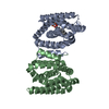


 PDBj
PDBj Assembly


 Mass: 22.990 Da / Num. of mol.: 2 / Source method: obtained synthetically / Formula: Na
 Mass: 18.015 Da / Num. of mol.: 122 / Source method: isolated from a natural source / Formula: H2O
 Sample preparation
 / Beamline: 19-ID / Wavelength: 0.97929 Å
 Processing