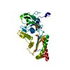
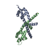
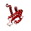
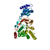
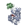
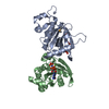
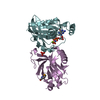
Entry Database : PDB / ID : 3a58Title Crystal structure of Sec3p - Rho1p complex from Saccharomyces cerevisiae Exocyst complex component SEC3 GTP-binding protein RHO1 Keywords / / / / / / / / / / / / / / / / / / Function / homology Function Domain/homology Component
/ / / / / / / / / / / / / / / / / / / / / / / / / / / / / / / / / / / / / / / / / / / / / / / / / / / / / / / / / / / / / / / / / / / / / / / / / / / / / / / / / / / / / / / / / / / / / / / / / / / / / / / / / / / / / / / / Biological species Saccharomyces cerevisiae (brewer's yeast)Method / / / Resolution : 2.6 Å Authors Yamashita, M. / Sato, Y. / Yamagata, A. / Mimura, H. / Yoshikawa, A. / Fukai, S. Journal : Nat.Struct.Mol.Biol. / Year : 2010Title : Structural basis for the Rho- and phosphoinositide-dependent localization of the exocyst subunit Sec3Authors : Yamashita, M. / Kurokawa, K. / Sato, Y. / Yamagata, A. / Mimura, H. / Yoshikawa, A. / Sato, K. / Nakano, A. / Fukai, S. History Deposition Aug 3, 2009 Deposition site / Processing site Revision 1.0 Jan 12, 2010 Provider / Type Revision 1.1 Jul 13, 2011 Group Revision 1.2 Sep 18, 2013 Group Revision 1.3 Nov 10, 2021 Group / Derived calculationsCategory database_2 / pdbx_struct_conn_angle ... database_2 / pdbx_struct_conn_angle / struct_conn / struct_ref_seq_dif / struct_site Item _database_2.pdbx_DOI / _database_2.pdbx_database_accession ... _database_2.pdbx_DOI / _database_2.pdbx_database_accession / _pdbx_struct_conn_angle.ptnr1_auth_comp_id / _pdbx_struct_conn_angle.ptnr1_auth_seq_id / _pdbx_struct_conn_angle.ptnr1_label_asym_id / _pdbx_struct_conn_angle.ptnr1_label_atom_id / _pdbx_struct_conn_angle.ptnr1_label_comp_id / _pdbx_struct_conn_angle.ptnr1_label_seq_id / _pdbx_struct_conn_angle.ptnr3_auth_comp_id / _pdbx_struct_conn_angle.ptnr3_auth_seq_id / _pdbx_struct_conn_angle.ptnr3_label_asym_id / _pdbx_struct_conn_angle.ptnr3_label_atom_id / _pdbx_struct_conn_angle.ptnr3_label_comp_id / _pdbx_struct_conn_angle.ptnr3_label_seq_id / _pdbx_struct_conn_angle.value / _struct_conn.pdbx_dist_value / _struct_conn.pdbx_leaving_atom_flag / _struct_conn.ptnr1_auth_asym_id / _struct_conn.ptnr1_auth_comp_id / _struct_conn.ptnr1_auth_seq_id / _struct_conn.ptnr1_label_asym_id / _struct_conn.ptnr1_label_atom_id / _struct_conn.ptnr1_label_comp_id / _struct_conn.ptnr1_label_seq_id / _struct_conn.ptnr2_auth_asym_id / _struct_conn.ptnr2_auth_comp_id / _struct_conn.ptnr2_auth_seq_id / _struct_conn.ptnr2_label_asym_id / _struct_conn.ptnr2_label_atom_id / _struct_conn.ptnr2_label_comp_id / _struct_ref_seq_dif.details / _struct_site.pdbx_auth_asym_id / _struct_site.pdbx_auth_comp_id / _struct_site.pdbx_auth_seq_id Revision 1.4 Oct 23, 2024 Group / Structure summaryCategory chem_comp_atom / chem_comp_bond ... chem_comp_atom / chem_comp_bond / pdbx_entry_details / pdbx_modification_feature
Show all Show less
 Movie
Movie Controller

 Yorodumi
 Open data
 Basic information
 Components
 Keywords
 Function and homology information
 X-RAY DIFFRACTION /
  Authors
 Citation
 Structure visualization
 Downloads & links
 Other downloads
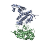

 PDBj
PDBj


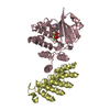
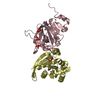
 Assembly
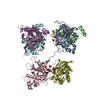


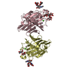
 Mass: 94.971 Da / Num. of mol.: 7 / Source method: obtained synthetically / Formula: PO4
 Mass: 522.196 Da / Num. of mol.: 3 / Source method: obtained synthetically / Formula: C10H17N6O13P3
 Mass: 24.305 Da / Num. of mol.: 3 / Source method: obtained synthetically / Formula: Mg
 Sample preparation
 / Beamline: BL41XU / Wavelength: 0.97924 Å
 Processing