 Movie
Movie Controller

[English] 日本語
 Yorodumi
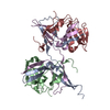
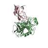
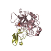
- PDB-3nfh: Crystal structure of tandem winged helix domain of RNA polymerase... -
+ Open data

- Basic information
| Entry | Database: PDB / ID: 3nfh | ||||||
|---|---|---|---|---|---|---|---|
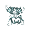
| Title | Crystal structure of tandem winged helix domain of RNA polymerase I subunit A49 (P4) | ||||||
 Components Components | DNA-directed RNA polymerase I subunit RPA49 | ||||||
 Keywords Keywords | TRANSCRIPTION / DNA BINDING PROTEIN / winged helix / RNA polymerase / DNA binding | ||||||
| Function / homology |  Function and homology information Function and homology informationRNA polymerase I preinitiation complex assembly / RNA Polymerase I Transcription Initiation / regulation of cell size / RNA Polymerase I Promoter Escape / termination of RNA polymerase I transcription / nucleolar large rRNA transcription by RNA polymerase I / transcription initiation at RNA polymerase I promoter / RNA polymerase I complex / transcription elongation by RNA polymerase I / transcription by RNA polymerase I ...RNA polymerase I preinitiation complex assembly / RNA Polymerase I Transcription Initiation / regulation of cell size / RNA Polymerase I Promoter Escape / termination of RNA polymerase I transcription / nucleolar large rRNA transcription by RNA polymerase I / transcription initiation at RNA polymerase I promoter / RNA polymerase I complex / transcription elongation by RNA polymerase I / transcription by RNA polymerase I / DNA-directed RNA polymerase activity / ribosome biogenesis / nucleolus / DNA binding / nucleus Similarity search - Function | ||||||
| Biological species |  | ||||||
| Method |  X-RAY DIFFRACTION / SYNCHROTRON / MOLECULAR REPLACEMENT / Resolution: 2.17 Å X-RAY DIFFRACTION / SYNCHROTRON / MOLECULAR REPLACEMENT / Resolution: 2.17 Å | ||||||
 Authors Authors | Geiger, S.R. / Lorenzen, K. / Schreieck, A. / Hanecker, P. / Kostrewa, D. / Heck, A.J.R. / Cramer, P. | ||||||
 Citation Citation | Journal: Mol.Cell / Year: 2010 Title: RNA Polymerase I Contains a TFIIF-Related DNA-Binding Subcomplex. Authors: Geiger, S.R. / Lorenzen, K. / Schreieck, A. / Hanecker, P. / Kostrewa, D. / Heck, A.J. / Cramer, P. | ||||||
| History |
|
- Structure visualization
| Structure viewer | Molecule: MolmilJmol/JSmol |
|---|
- Downloads & links
-Download
| PDBx/mmCIF format | 3nfh.cif.gz | 184.1 KB | Display | PDBx/mmCIF format |
|---|---|---|---|---|
| PDB format | pdb3nfh.ent.gz | 147.6 KB | Display | PDB format |
| PDBx/mmJSON format | 3nfh.json.gz | Tree view | PDBx/mmJSON format | |
| Others |  Other downloads Other downloads |
-Validation report
| Arichive directory | https://data.pdbj.org/pub/pdb/validation_reports/nf/3nfhftp://data.pdbj.org/pub/pdb/validation_reports/nf/3nfh | HTTPS FTP |
|---|
-Related structure data


-Links
 PDBj
PDBj


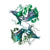
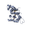
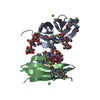
- Assembly
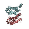
| Deposited unit | 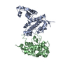
| ||||||||
|---|---|---|---|---|---|---|---|---|---|
| 1 | 
| ||||||||
| 2 | 
| ||||||||
| Unit cell |
|
-Components
| #1: Protein | Mass: 27717.176 Da / Num. of mol.: 2 / Fragment: UNP residues 154-399 Source method: isolated from a genetically manipulated source Source: (gene. exp.) Gene: N0880, RPA49, RRN13, YNL248C / Plasmid: pET28b / Production host:  #2: Water | ChemComp-HOH / |  Mass: 18.015 Da / Num. of mol.: 205 / Source method: isolated from a natural source / Formula: H2O Mass: 18.015 Da / Num. of mol.: 205 / Source method: isolated from a natural source / Formula: H2O |
|---|
-Experimental details
-Experiment
| Experiment | Method: X-RAY DIFFRACTION / Number of used crystals: 1 |
|---|
- Sample preparation
| Crystal | Density Matthews: 2.3 Å3/Da / Density % sol: 46.6 % |
|---|---|
| Crystal grow | Temperature: 277 K / Method: vapor diffusion, hanging drop / pH: 8.5 Details: 26% PEG 4000, 200 mM lithium sulfate monohydrate, 100 mM Tris pH 8.5, VAPOR DIFFUSION, HANGING DROP, temperature 277K |
-Data collection
| Diffraction | Mean temperature: 70 K |
|---|---|
| Diffraction source | Source: SYNCHROTRON / Site: ESRF  / Beamline: ID14-4 / Wavelength: 0.979 Å / Beamline: ID14-4 / Wavelength: 0.979 Å |
| Detector | Type: ADSC QUANTUM 315r / Detector: CCD / Date: May 1, 2009 / Details: torodial focusing mirror |
| Radiation | Monochromator: channel cut ESRF monochromator / Protocol: SINGLE WAVELENGTH / Monochromatic (M) / Laue (L): M / Scattering type: x-ray |
| Radiation wavelength | Wavelength: 0.979 Å / Relative weight: 1 |
| Reflection | Resolution: 2.17→80 Å / Num. obs: 26581 / % possible obs: 99.8 % / Observed criterion σ(F): 0 / Observed criterion σ(I): 0 / Redundancy: 11.7 % / Biso Wilson estimate: 30.32 Å2 / Rmerge(I) obs: 0.08 / Rsym value: 0.08 / Net I/σ(I): 27.5 |
| Reflection shell | Resolution: 2.17→2.45 Å / Redundancy: 12.5 % / Rmerge(I) obs: 0.597 / Mean I/σ(I) obs: 6.3 / Rsym value: 0.597 / % possible all: 99.8 |
- Processing
| Software |
| ||||||||||||||||||||||||||||||||||||||||||||||||||||||||||||||||||||||||||||||||||||||||||||||||||||||||||||||||||
|---|---|---|---|---|---|---|---|---|---|---|---|---|---|---|---|---|---|---|---|---|---|---|---|---|---|---|---|---|---|---|---|---|---|---|---|---|---|---|---|---|---|---|---|---|---|---|---|---|---|---|---|---|---|---|---|---|---|---|---|---|---|---|---|---|---|---|---|---|---|---|---|---|---|---|---|---|---|---|---|---|---|---|---|---|---|---|---|---|---|---|---|---|---|---|---|---|---|---|---|---|---|---|---|---|---|---|---|---|---|---|---|---|---|---|---|
| Refinement | Method to determine structure: MOLECULAR REPLACEMENT Starting model: Crystal structure of tandem winged helix domain of RNA polymerase I subunit A49 (SG P21, on hold) Resolution: 2.17→36.2 Å / Cor.coef. Fo:Fc: 0.9433 / Cor.coef. Fo:Fc free: 0.9162 / Cross valid method: THROUGHOUT / σ(F): 0 / Stereochemistry target values: Engh & Huber
| ||||||||||||||||||||||||||||||||||||||||||||||||||||||||||||||||||||||||||||||||||||||||||||||||||||||||||||||||||
| Displacement parameters | Biso mean: 40.39 Å2
| ||||||||||||||||||||||||||||||||||||||||||||||||||||||||||||||||||||||||||||||||||||||||||||||||||||||||||||||||||
| Refine analyze | Luzzati coordinate error obs: 0.292 Å | ||||||||||||||||||||||||||||||||||||||||||||||||||||||||||||||||||||||||||||||||||||||||||||||||||||||||||||||||||
| Refinement step | Cycle: LAST / Resolution: 2.17→36.2 Å
| ||||||||||||||||||||||||||||||||||||||||||||||||||||||||||||||||||||||||||||||||||||||||||||||||||||||||||||||||||
| Refine LS restraints |
| ||||||||||||||||||||||||||||||||||||||||||||||||||||||||||||||||||||||||||||||||||||||||||||||||||||||||||||||||||
| LS refinement shell | Resolution: 2.17→2.26 Å / Total num. of bins used: 13
| ||||||||||||||||||||||||||||||||||||||||||||||||||||||||||||||||||||||||||||||||||||||||||||||||||||||||||||||||||
| Refinement TLS params. | Method: refined / Refine-ID: X-RAY DIFFRACTION
| ||||||||||||||||||||||||||||||||||||||||||||||||||||||||||||||||||||||||||||||||||||||||||||||||||||||||||||||||||
| Refinement TLS group |
|
