 Movie
Movie Controller


+ Open data

- Basic information
| Entry | Database: PDB / ID: 4gec | ||||||
|---|---|---|---|---|---|---|---|
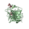
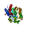
| Title | Crystal Structure of E.coli MenH R124A Mutant | ||||||
 Components Components | 2-succinyl-6-hydroxy-2,4-cyclohexadiene-1-carboxylate synthase | ||||||
 Keywords Keywords | LYASE / menaquinone biosynthesis / alpha beta hydrolase / 2-succinyl-6-hydroxy-2 / 4-cyclohexadiene-1-carboxylate synthase / mutant R124A / MenH mutant R124A | ||||||
| Function / homology |  Function and homology information Function and homology information2-succinyl-6-hydroxy-2,4-cyclohexadiene-1-carboxylate synthase / 2-succinyl-6-hydroxy-2,4-cyclohexadiene-1-carboxylate synthase activity / menaquinone biosynthetic process / cytosol Similarity search - Function | ||||||
| Biological species |  | ||||||
| Method |  X-RAY DIFFRACTION / SYNCHROTRON / MOLECULAR REPLACEMENT / molecular replacement / Resolution: 2.5 Å X-RAY DIFFRACTION / SYNCHROTRON / MOLECULAR REPLACEMENT / molecular replacement / Resolution: 2.5 Å | ||||||
 Authors Authors | Johnston, J.M. / Baker, E.N. / Guo, Z. / Jiang, M. | ||||||
 Citation Citation | Journal: Plos One / Year: 2013 Title: Crystal Structures of E. coli Native MenH and Two Active Site Mutants. Authors: Johnston, J.M. / Jiang, M. / Guo, Z. / Baker, E.N. | ||||||
| History |
|
- Structure visualization
| Structure viewer | Molecule: MolmilJmol/JSmol |
|---|
- Downloads & links
-Download
| PDBx/mmCIF format | 4gec.cif.gz | 162.3 KB | Display | PDBx/mmCIF format |
|---|---|---|---|---|
| PDB format | pdb4gec.ent.gz | 128.2 KB | Display | PDB format |
| PDBx/mmJSON format | 4gec.json.gz | Tree view | PDBx/mmJSON format | |
| Others |  Other downloads Other downloads |
-Validation report
| Arichive directory | https://data.pdbj.org/pub/pdb/validation_reports/ge/4gecftp://data.pdbj.org/pub/pdb/validation_reports/ge/4gec | HTTPS FTP |
|---|
-Related structure data


-Links
 PDBj
PDBj

- Assembly
| Deposited unit | 
| ||||||||
|---|---|---|---|---|---|---|---|---|---|
| 1 | 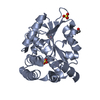
| ||||||||
| 2 | 
| ||||||||
| 3 | 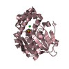
| ||||||||
| Unit cell |
|
-Components
-Protein , 1 types, 3 molecules ABC
| #1: Protein | Mass: 29429.234 Da / Num. of mol.: 3 / Mutation: R124A Source method: isolated from a genetically manipulated source Source: (gene. exp.) References: UniProt: P37355, 2-succinyl-6-hydroxy-2,4-cyclohexadiene-1-carboxylate synthase |
|---|
-Non-polymers , 5 types, 241 molecules 


| #2: Chemical | ChemComp-SO4 /  Mass: 96.063 Da / Num. of mol.: 4 / Source method: obtained synthetically / Formula: SO4 Mass: 96.063 Da / Num. of mol.: 4 / Source method: obtained synthetically / Formula: SO4#3: Chemical | ChemComp-GOL / |  Mass: 92.094 Da / Num. of mol.: 1 / Source method: obtained synthetically / Formula: C3H8O3 Mass: 92.094 Da / Num. of mol.: 1 / Source method: obtained synthetically / Formula: C3H8O3#4: Chemical |  Mass: 35.453 Da / Num. of mol.: 3 / Source method: obtained synthetically / Formula: Cl Mass: 35.453 Da / Num. of mol.: 3 / Source method: obtained synthetically / Formula: Cl#5: Chemical | ChemComp-EDO / |  Mass: 62.068 Da / Num. of mol.: 1 / Source method: obtained synthetically / Formula: C2H6O2 Mass: 62.068 Da / Num. of mol.: 1 / Source method: obtained synthetically / Formula: C2H6O2#6: Water | ChemComp-HOH / | Mass: 18.015 Da / Num. of mol.: 232 / Source method: isolated from a natural source / Formula: H2O |
|---|
-Experimental details
-Experiment
| Experiment | Method: X-RAY DIFFRACTION / Number of used crystals: 1 |
|---|
- Sample preparation
| Crystal | Density Matthews: 4.41 Å3/Da / Density % sol: 72.1 % |
|---|---|
| Crystal grow | Temperature: 291 K / Method: vapor diffusion, hanging drop / pH: 5.5 Details: Sodium Citrate, Ammonium sulfate, Lithium sulfate, pH 5.5, vapor diffusion, hanging drop, temperature 291K |
-Data collection
| Diffraction | Mean temperature: 110 K | ||||||||||||||||||||||||||||||||||||||||||||||||||||||||||||||||||||||||||||||||||||||||
|---|---|---|---|---|---|---|---|---|---|---|---|---|---|---|---|---|---|---|---|---|---|---|---|---|---|---|---|---|---|---|---|---|---|---|---|---|---|---|---|---|---|---|---|---|---|---|---|---|---|---|---|---|---|---|---|---|---|---|---|---|---|---|---|---|---|---|---|---|---|---|---|---|---|---|---|---|---|---|---|---|---|---|---|---|---|---|---|---|---|
| Diffraction source | Source: SYNCHROTRON / Site: Australian Synchrotron  / Beamline: MX1 / Wavelength: 0.953692 Å / Beamline: MX1 / Wavelength: 0.953692 Å | ||||||||||||||||||||||||||||||||||||||||||||||||||||||||||||||||||||||||||||||||||||||||
| Detector | Type: ADSC QUANTUM 210r / Detector: CCD / Date: Dec 15, 2010 | ||||||||||||||||||||||||||||||||||||||||||||||||||||||||||||||||||||||||||||||||||||||||
| Radiation | Protocol: SINGLE WAVELENGTH / Monochromatic (M) / Laue (L): M / Scattering type: x-ray | ||||||||||||||||||||||||||||||||||||||||||||||||||||||||||||||||||||||||||||||||||||||||
| Radiation wavelength | Wavelength: 0.953692 Å / Relative weight: 1 | ||||||||||||||||||||||||||||||||||||||||||||||||||||||||||||||||||||||||||||||||||||||||
| Reflection | Resolution: 2.5→117.444 Å / Num. all: 54795 / Num. obs: 54795 / % possible obs: 99.4 % / Redundancy: 14.9 % / Biso Wilson estimate: 61.48 Å2 / Rmerge(I) obs: 0.19 / Rsym value: 0.19 / Net I/σ(I): 17.6 | ||||||||||||||||||||||||||||||||||||||||||||||||||||||||||||||||||||||||||||||||||||||||
| Reflection shell | Diffraction-ID: 1
|
-Phasing
| Phasing | Method: molecular replacement |
|---|
- Processing
| Software |
| ||||||||||||||||||||||||||||||||||||||||||||||||||||||||||||||||||||||||||||||||||||||||||||||||||||||||||||
|---|---|---|---|---|---|---|---|---|---|---|---|---|---|---|---|---|---|---|---|---|---|---|---|---|---|---|---|---|---|---|---|---|---|---|---|---|---|---|---|---|---|---|---|---|---|---|---|---|---|---|---|---|---|---|---|---|---|---|---|---|---|---|---|---|---|---|---|---|---|---|---|---|---|---|---|---|---|---|---|---|---|---|---|---|---|---|---|---|---|---|---|---|---|---|---|---|---|---|---|---|---|---|---|---|---|---|---|---|---|
| Refinement | Method to determine structure: MOLECULAR REPLACEMENT / Resolution: 2.5→81.42 Å / Cor.coef. Fo:Fc: 0.9363 / Cor.coef. Fo:Fc free: 0.922 / Occupancy max: 1 / Occupancy min: 1 / SU R Cruickshank DPI: 0.22 / Cross valid method: THROUGHOUT / σ(F): 0 / SU R Blow DPI: 0.223 / SU Rfree Blow DPI: 0.187 / SU Rfree Cruickshank DPI: 0.187 / Stereochemistry target values: Engh & Huber
| ||||||||||||||||||||||||||||||||||||||||||||||||||||||||||||||||||||||||||||||||||||||||||||||||||||||||||||
| Displacement parameters | Biso max: 141.12 Å2 / Biso mean: 49.133 Å2 / Biso min: 17.7 Å2
| ||||||||||||||||||||||||||||||||||||||||||||||||||||||||||||||||||||||||||||||||||||||||||||||||||||||||||||
| Refine analyze | Luzzati coordinate error obs: 0.322 Å | ||||||||||||||||||||||||||||||||||||||||||||||||||||||||||||||||||||||||||||||||||||||||||||||||||||||||||||
| Refinement step | Cycle: LAST / Resolution: 2.5→81.42 Å
| ||||||||||||||||||||||||||||||||||||||||||||||||||||||||||||||||||||||||||||||||||||||||||||||||||||||||||||
| Refine LS restraints |
| ||||||||||||||||||||||||||||||||||||||||||||||||||||||||||||||||||||||||||||||||||||||||||||||||||||||||||||
| LS refinement shell | Resolution: 2.5→2.56 Å / Total num. of bins used: 20
|
