 Movie
Movie Controller


+ Open data

- Basic information
| Entry | Database: PDB / ID: 4g14 | ||||||
|---|---|---|---|---|---|---|---|
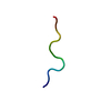
| Title | Crystal structure of samarosporin I at 293K | ||||||
 Components Components | SAMAROSPORIN I | ||||||
 Keywords Keywords | ANTIBIOTIC / peptaibol / 3(10)-alpha Helix / antibiotic peptide / membrane / extracellular | ||||||
| Function / homology | SAMAROSPORIN I / :  Function and homology information Function and homology information | ||||||
| Biological species | samarospora rostrup (unknown) | ||||||
| Method |  X-RAY DIFFRACTION / SYNCHROTRON / MOLECULAR REPLACEMENT / Resolution: 1.09 Å X-RAY DIFFRACTION / SYNCHROTRON / MOLECULAR REPLACEMENT / Resolution: 1.09 Å | ||||||
 Authors Authors | Gessmann, R. / Axford, D. / Petratos, K. | ||||||
 Citation Citation | Journal: J.Pept.Sci. / Year: 2012 Title: The crystal structure of samarosporin I at atomic resolution. Authors: Gessmann, R. / Axford, D. / Evans, G. / Bruckner, H. / Petratos, K. | ||||||
| History |
|
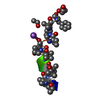
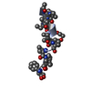
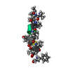
- Structure visualization
| Structure viewer | Molecule: MolmilJmol/JSmol |
|---|
- Downloads & links
-Download
| PDBx/mmCIF format | 4g14.cif.gz | 17.7 KB | Display | PDBx/mmCIF format |
|---|---|---|---|---|
| PDB format | pdb4g14.ent.gz | 12.1 KB | Display | PDB format |
| PDBx/mmJSON format | 4g14.json.gz | Tree view | PDBx/mmJSON format | |
| Others |  Other downloads Other downloads |
-Validation report
| Summary document | 4g14_validation.pdf.gz | 408.5 KB | Display | wwPDB validaton report |
|---|---|---|---|---|
| Full document | 4g14_full_validation.pdf.gz | 408.5 KB | Display | |
| Data in XML | 4g14_validation.xml.gz | 3.1 KB | Display | |
| Data in CIF | 4g14_validation.cif.gz | 3.3 KB | Display | |
| Arichive directory | https://data.pdbj.org/pub/pdb/validation_reports/g1/4g14ftp://data.pdbj.org/pub/pdb/validation_reports/g1/4g14 | HTTPS FTP |
-Related structure data
| Related structure data | 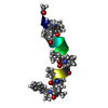 4g13SC S: Starting model for refinement C: citing same article ( |
|---|---|
| Similar structure data |


-Links
 PDBj
PDBj- Assembly
| Deposited unit | 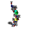
| ||||||||
|---|---|---|---|---|---|---|---|---|---|
| 1 |
| ||||||||
| Unit cell |
| ||||||||
| Details | AS PER AUTHORS THE BIOLOGICAL ASSEMBLY IS MULTIMERIC OF UNKNOWN STOICHIOMETRY |
-Components
| #1: Protein/peptide | 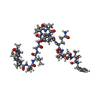  Type: Peptaibol / Class: Antibiotic / Mass: 1557.876 Da / Num. of mol.: 1 / Source method: isolated from a natural source / Source: (natural) samarospora rostrup (unknown) / Strain: F-7762 / References: NOR: NOR00986, SAMAROSPORIN I Type: Peptaibol / Class: Antibiotic / Mass: 1557.876 Da / Num. of mol.: 1 / Source method: isolated from a natural source / Source: (natural) samarospora rostrup (unknown) / Strain: F-7762 / References: NOR: NOR00986, SAMAROSPORIN I |
|---|---|
| #2: Water | ChemComp-HOH /  Mass: 18.015 Da / Num. of mol.: 2 / Source method: isolated from a natural source / Formula: H2O Mass: 18.015 Da / Num. of mol.: 2 / Source method: isolated from a natural source / Formula: H2O |
| Compound details | SAMAROSPORIN I/EMERIMICIN IV IS LINEAR PEPTIDE, A MEMBER OF THE PEPT FAMILY OF MEMBRANE CHANNEL ...SAMAROSPOR |
-Experimental details
-Experiment
| Experiment | Method: X-RAY DIFFRACTION / Number of used crystals: 1 |
|---|
- Sample preparation
| Crystal | Density Matthews: 1.52 Å3/Da / Density % sol: 18.4 % |
|---|---|
| Crystal grow | Temperature: 293 K / Method: evaporation / Details: methanol/water, EVAPORATION, temperature 293K |
-Data collection
| Diffraction | Mean temperature: 293 K |
|---|---|
| Diffraction source | Source: SYNCHROTRON / Site: Diamond  / Beamline: I24 / Wavelength: 0.7293 Å / Beamline: I24 / Wavelength: 0.7293 Å |
| Detector | Type: DECTRIS PILATUS 6M / Detector: PIXEL / Date: Mar 3, 2012 |
| Radiation | Monochromator: ACCEL FIXED EXIT DOUBLE CRYSTAL / Protocol: SINGLE WAVELENGTH / Monochromatic (M) / Laue (L): M / Scattering type: x-ray |
| Radiation wavelength | Wavelength: 0.7293 Å / Relative weight: 1 |
| Reflection | Resolution: 1.09→22.59 Å / Num. all: 4194 / Num. obs: 4194 / % possible obs: 99.2 % / Observed criterion σ(F): 0 / Observed criterion σ(I): 0 / Redundancy: 3 % / Net I/σ(I): 4.2 |
| Reflection shell | Resolution: 1.09→1.12 Å / Redundancy: 3.1 % / Mean I/σ(I) obs: 2.6 / % possible all: 99.2 |
- Processing
| Software |
| |||||||||||||||||||||||||
|---|---|---|---|---|---|---|---|---|---|---|---|---|---|---|---|---|---|---|---|---|---|---|---|---|---|---|
| Refinement | Method to determine structure: MOLECULAR REPLACEMENT Starting model: PDB ENTRY 4G13 Resolution: 1.09→22.59 Å / Num. parameters: 1022 / Num. restraintsaints: 1001 / σ(F): 0 / Stereochemistry target values: Engh & Huber
| |||||||||||||||||||||||||
| Solvent computation | Solvent model: BABINET | |||||||||||||||||||||||||
| Refine analyze | Occupancy sum hydrogen: 112 / Occupancy sum non hydrogen: 114 | |||||||||||||||||||||||||
| Refinement step | Cycle: LAST / Resolution: 1.09→22.59 Å
|
