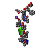
 Movie
Movie Controller


+ Open data

- Basic information
| Entry | Database: PDB / ID: 1ob4 | ||||||
|---|---|---|---|---|---|---|---|
| Title | Cephaibol A | ||||||
 Components Components | CEPHAIBOL A | ||||||
 Keywords Keywords | ANTIBIOTIC / ION CHANNEL / CEPHAIBOL / PEPTAIBOL / ANTIBACTERIAL / ANTIFUNGAL | ||||||
| Function / homology | Cephaibol A / ETHANOL / :  Function and homology information Function and homology information | ||||||
| Biological species |  ACREMONIUM TUBAKII (fungus) ACREMONIUM TUBAKII (fungus) | ||||||
| Method |  X-RAY DIFFRACTION / DIRECT METHODS / Resolution: 0.95 Å X-RAY DIFFRACTION / DIRECT METHODS / Resolution: 0.95 Å | ||||||
 Authors Authors | Bunkoczi, G. / Schiell, M. / Vertesy, L. / Sheldrick, G.M. | ||||||
 Citation Citation | Journal: J.Pept.Sci. / Year: 2003 Title: Crystal Structures of Cephaibols Authors: Bunkoczi, G. / Schiell, M. / Vertesy, L. / Sheldrick, G.M. | ||||||
| History |
|
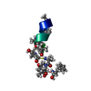
- Structure visualization
| Structure viewer | Molecule: MolmilJmol/JSmol |
|---|
- Downloads & links
-Download
| PDBx/mmCIF format | 1ob4.cif.gz | 17.9 KB | Display | PDBx/mmCIF format |
|---|---|---|---|---|
| PDB format | pdb1ob4.ent.gz | 11.3 KB | Display | PDB format |
| PDBx/mmJSON format | 1ob4.json.gz | Tree view | PDBx/mmJSON format | |
| Others |  Other downloads Other downloads |
-Validation report
| Arichive directory | https://data.pdbj.org/pub/pdb/validation_reports/ob/1ob4ftp://data.pdbj.org/pub/pdb/validation_reports/ob/1ob4 | HTTPS FTP |
|---|
-Related structure data


-Links
 PDBj
PDBj


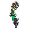
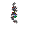
- Assembly
| Deposited unit | 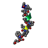
| ||||||||
|---|---|---|---|---|---|---|---|---|---|
| 1 |
| ||||||||
| Unit cell |
|
-Components
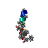
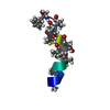
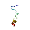
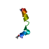
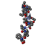
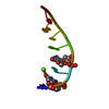
| #1: Protein/peptide |   Type: Peptaibol / Class: Antibiotic / Mass: 1654.991 Da / Num. of mol.: 1 / Source method: isolated from a natural source Type: Peptaibol / Class: Antibiotic / Mass: 1654.991 Da / Num. of mol.: 1 / Source method: isolated from a natural sourceDetails: CEPHAIBOL A IS A HEXADECAMERIC HELICAL PEPTIDE. THE N-TERM IS ACETYLATED (RESIDUE 0) Source: (natural) ACREMONIUM TUBAKII (fungus) / References: NOR: NOR00969, Cephaibol A |
|---|---|
| #2: Chemical | ChemComp-EOH /   Mass: 46.068 Da / Num. of mol.: 1 / Source method: obtained synthetically / Formula: C2H6O Mass: 46.068 Da / Num. of mol.: 1 / Source method: obtained synthetically / Formula: C2H6O |
| #3: Water | ChemComp-HOH /  Mass: 18.015 Da / Num. of mol.: 15 / Source method: isolated from a natural source / Formula: H2O Mass: 18.015 Da / Num. of mol.: 15 / Source method: isolated from a natural source / Formula: H2O |
| Compound details | CEPHAIBOL A IS LINEAR PEPTIDE, A MEMBER OF THE PEPTAIBOL FAMILY OF MEMBRANE CHANNEL FORMING ...CEPHAIBOL A IS LINEAR PEPTIDE, A MEMBER OF THE PEPTAIBOL FAMILY OF MEMBRANE CHANNEL FORMING PEPTIDES. HERE, CEPHAIBOL A IS REPRESENTE |
| Has protein modification | Y |
-Experimental details
-Experiment
| Experiment | Method: X-RAY DIFFRACTION / Number of used crystals: 1 |
|---|
- Sample preparation
| Crystal | Density Matthews: 1.57 Å3/Da / Density % sol: 22 % | ||||||||||||||||||||||||
|---|---|---|---|---|---|---|---|---|---|---|---|---|---|---|---|---|---|---|---|---|---|---|---|---|---|
| Crystal grow | pH: 4.6 / Details: 0.1 M NAAC/HAC PH=4.6, 34% ETOH, pH 4.60 | ||||||||||||||||||||||||
| Crystal grow | *PLUS Method: vapor diffusion, hanging drop / PH range low: 4.8 / PH range high: 4.2 | ||||||||||||||||||||||||
| Components of the solutions | *PLUS
|
-Data collection
| Diffraction | Mean temperature: 100 K |
|---|---|
| Diffraction source | Source: ROTATING ANODE / Type: BRUKER M06XCE / Wavelength: 1.5418 |
| Detector | Type: BRUKER SMART 6000 / Detector: CCD / Date: Feb 15, 2002 / Details: MIRRORS |
| Radiation | Protocol: SINGLE WAVELENGTH / Monochromatic (M) / Laue (L): M / Scattering type: x-ray |
| Radiation wavelength | Wavelength: 1.5418 Å / Relative weight: 1 |
| Reflection | Resolution: 0.95→37.8 Å / Num. obs: 7025 / % possible obs: 98.6 % / Redundancy: 9.45 % / Rmerge(I) obs: 0.0431 / Net I/σ(I): 29.21 |
| Reflection shell | Resolution: 0.95→1.05 Å / Redundancy: 4.66 % / Rmerge(I) obs: 0.1156 / Mean I/σ(I) obs: 10.67 / % possible all: 96.6 |
| Reflection | *PLUS Highest resolution: 0.95 Å / Redundancy: 9.45 % / Num. measured all: 67303 / Rmerge(I) obs: 0.0431 |
| Reflection shell | *PLUS % possible obs: 96.6 % / Rmerge(I) obs: 0.1156 / Mean I/σ(I) obs: 10.67 |
- Processing
| Software |
| |||||||||||||||||||||||||||||||||
|---|---|---|---|---|---|---|---|---|---|---|---|---|---|---|---|---|---|---|---|---|---|---|---|---|---|---|---|---|---|---|---|---|---|---|
| Refinement | Method to determine structure: DIRECT METHODS / Resolution: 0.95→37.8 Å / Num. parameters: 1429 / Num. restraintsaints: 2016 / Cross valid method: FREE R-VALUE / σ(F): 0 StereochEM target val spec case: RESTRAINTS FOR AIB, DIV, HYP AND PHL FROM ANTIAMOEBIN (PDB 1JOH) Stereochemistry target values: ENGH AND HUBER
| |||||||||||||||||||||||||||||||||
| Solvent computation | Solvent model: MOEWS & KRETSINGER | |||||||||||||||||||||||||||||||||
| Refine analyze | Num. disordered residues: 2 / Occupancy sum hydrogen: 124 / Occupancy sum non hydrogen: 133.28 | |||||||||||||||||||||||||||||||||
| Refinement step | Cycle: LAST / Resolution: 0.95→37.8 Å
| |||||||||||||||||||||||||||||||||
| Refine LS restraints |
| |||||||||||||||||||||||||||||||||
| Software | *PLUS Name: SHELXL / Version: 97 / Classification: refinement | |||||||||||||||||||||||||||||||||
| Refinement | *PLUS Lowest resolution: 37.79 Å / Rfactor Rwork: 0.0872 | |||||||||||||||||||||||||||||||||
| Solvent computation | *PLUS | |||||||||||||||||||||||||||||||||
| Displacement parameters | *PLUS | |||||||||||||||||||||||||||||||||
| Refine LS restraints | *PLUS
|
