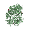
 Movie
Movie Controller


+ Open data

- Basic information
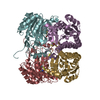
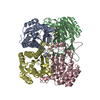
| Entry | Database: PDB / ID: 4fxd | ||||||
|---|---|---|---|---|---|---|---|
| Title | Crystal structure of yeast DNA polymerase alpha bound to DNA/RNA | ||||||
 Components Components |
| ||||||
 Keywords Keywords | TRANSFERASE/DNA/RNA / DNA polymerase / DNA replication / TRANSFERASE-DNA-RNA complex | ||||||
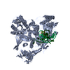
| Function / homology |  Function and homology information Function and homology informationInhibition of replication initiation of damaged DNA by RB1/E2F1 / H3-H4 histone complex chaperone activity / DNA replication initiation / Processive synthesis on the lagging strand / RNA-templated DNA biosynthetic process / Removal of the Flap Intermediate / Polymerase switching / premeiotic DNA replication / alpha DNA polymerase:primase complex / Activation of the pre-replicative complex ...Inhibition of replication initiation of damaged DNA by RB1/E2F1 / H3-H4 histone complex chaperone activity / DNA replication initiation / Processive synthesis on the lagging strand / RNA-templated DNA biosynthetic process / Removal of the Flap Intermediate / Polymerase switching / premeiotic DNA replication / alpha DNA polymerase:primase complex / Activation of the pre-replicative complex / lagging strand elongation / mitotic DNA replication initiation / leading strand elongation / DNA synthesis involved in DNA repair / DNA replication origin binding / DNA replication initiation / replication fork / double-strand break repair / single-stranded DNA binding / 4 iron, 4 sulfur cluster binding / DNA-directed DNA polymerase / DNA-directed DNA polymerase activity / DNA replication / nucleotide binding / chromatin binding / mitochondrion / zinc ion binding Similarity search - Function | ||||||
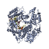
| Biological species |  | ||||||
| Method |  X-RAY DIFFRACTION / SYNCHROTRON / MOLECULAR REPLACEMENT / Resolution: 3 Å X-RAY DIFFRACTION / SYNCHROTRON / MOLECULAR REPLACEMENT / Resolution: 3 Å | ||||||
 Authors Authors | Perera, R.L. / Torella, R. / Klinge, S. / Kilkenny, M.L. / Maman, J.D. / Pellegrini, L. | ||||||
 Citation Citation | Journal: eLife / Year: 2013 Title: Mechanism for priming DNA synthesis by yeast DNA Polymerase alpha Authors: Perera, R.L. / Torella, R. / Klinge, S. / Kilkenny, M.L. / Maman, J.D. / Pellegrini, L. | ||||||
| History |
|
- Structure visualization
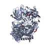
| Structure viewer | Molecule: MolmilJmol/JSmol |
|---|
- Downloads & links
-Download
| PDBx/mmCIF format | 4fxd.cif.gz | 659.8 KB | Display | PDBx/mmCIF format |
|---|---|---|---|---|
| PDB format | pdb4fxd.ent.gz | 540.8 KB | Display | PDB format |
| PDBx/mmJSON format | 4fxd.json.gz | Tree view | PDBx/mmJSON format | |
| Others |  Other downloads Other downloads |
-Validation report
| Arichive directory | https://data.pdbj.org/pub/pdb/validation_reports/fx/4fxdftp://data.pdbj.org/pub/pdb/validation_reports/fx/4fxd | HTTPS FTP |
|---|
-Related structure data
| Related structure data |  4b08C  4fvmSC  4fydC C: citing same article ( S: Starting model for refinement |
|---|---|
| Similar structure data |
-Links
 PDBj
PDBj


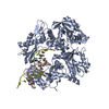
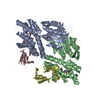
- Assembly
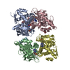
| Deposited unit | 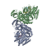
| ||||||||
|---|---|---|---|---|---|---|---|---|---|
| 1 | 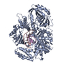
| ||||||||
| 2 | 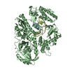
| ||||||||
| Unit cell |
|
-Components
| #1: Protein | Mass: 103531.828 Da / Num. of mol.: 2 / Fragment: Polymerase domain, UNP residues 349-1258 Source method: isolated from a genetically manipulated source Source: (gene. exp.) Strain: ATCC 204508 / S288c / Gene: CDC17, N2181, POL1, YNL102W / Plasmid: pRSFDuet-1 / Production host:  #2: DNA chain | Mass: 4792.089 Da / Num. of mol.: 2 / Source method: obtained synthetically / Details: Chemical synthesis #3: RNA chain | Mass: 3295.052 Da / Num. of mol.: 2 / Source method: obtained synthetically / Details: Chemical synthesis |
|---|
-Experimental details
-Experiment
| Experiment | Method: X-RAY DIFFRACTION / Number of used crystals: 1 |
|---|
- Sample preparation
| Crystal | Density Matthews: 2.66 Å3/Da / Density % sol: 53.75 % |
|---|---|
| Crystal grow | Temperature: 291 K / Method: vapor diffusion, hanging drop / pH: 8 Details: 0.1 M Bicine, 12% PEG3350, 10 mM MgCl2, pH 8.0, VAPOR DIFFUSION, HANGING DROP, temperature 291K |
-Data collection
| Diffraction | Mean temperature: 100 K |
|---|---|
| Diffraction source | Source: SYNCHROTRON / Site: ESRF  / Beamline: ID14-1 / Wavelength: 0.9334 Å / Beamline: ID14-1 / Wavelength: 0.9334 Å |
| Detector | Type: ADSC QUANTUM 210 / Detector: CCD / Date: Dec 11, 2009 |
| Radiation | Protocol: SINGLE WAVELENGTH / Monochromatic (M) / Laue (L): M / Scattering type: x-ray |
| Radiation wavelength | Wavelength: 0.9334 Å / Relative weight: 1 |
| Reflection | Resolution: 3→58.77 Å / Num. all: 45027 / Num. obs: 45027 / % possible obs: 97.8 % / Observed criterion σ(F): 0 / Observed criterion σ(I): 0 / Redundancy: 3.9 % / Biso Wilson estimate: 91.64 Å2 / Rmerge(I) obs: 0.073 / Rsym value: 0.073 / Net I/σ(I): 13.6 |
| Reflection shell | Resolution: 3→3.16 Å / Redundancy: 3.9 % / Rmerge(I) obs: 0.921 / Mean I/σ(I) obs: 1.5 / Num. unique all: 6546 / Rsym value: 0.921 / % possible all: 97.5 |
- Processing
| Software |
| |||||||||||||||||||||||||||||||||||||||||||||||||||||||||||||||||||||||||||||||||||||||||||||||||||||||||||||||||||||||
|---|---|---|---|---|---|---|---|---|---|---|---|---|---|---|---|---|---|---|---|---|---|---|---|---|---|---|---|---|---|---|---|---|---|---|---|---|---|---|---|---|---|---|---|---|---|---|---|---|---|---|---|---|---|---|---|---|---|---|---|---|---|---|---|---|---|---|---|---|---|---|---|---|---|---|---|---|---|---|---|---|---|---|---|---|---|---|---|---|---|---|---|---|---|---|---|---|---|---|---|---|---|---|---|---|---|---|---|---|---|---|---|---|---|---|---|---|---|---|---|---|
| Refinement | Method to determine structure: MOLECULAR REPLACEMENT Starting model: PDB ENTRY 4FVM Resolution: 3→55.526 Å / SU ML: 0.52 / Cross valid method: THROUGHOUT / σ(F): 0 / Phase error: 34.05 / Stereochemistry target values: ML Details: FOR REFINEMENT, REFMAC, BUSTER AND PHENIX WERE USED. THE STRUCTURE WAS REFINED WITH RIDING HYDROGENS. THE HYDROGENS OF THE LAST REFINEMENT RUN ARE INCLUDED.
| |||||||||||||||||||||||||||||||||||||||||||||||||||||||||||||||||||||||||||||||||||||||||||||||||||||||||||||||||||||||
| Solvent computation | Shrinkage radii: 0.8 Å / VDW probe radii: 1 Å / Solvent model: FLAT BULK SOLVENT MODEL. | |||||||||||||||||||||||||||||||||||||||||||||||||||||||||||||||||||||||||||||||||||||||||||||||||||||||||||||||||||||||
| Refinement step | Cycle: LAST / Resolution: 3→55.526 Å
| |||||||||||||||||||||||||||||||||||||||||||||||||||||||||||||||||||||||||||||||||||||||||||||||||||||||||||||||||||||||
| Refine LS restraints |
| |||||||||||||||||||||||||||||||||||||||||||||||||||||||||||||||||||||||||||||||||||||||||||||||||||||||||||||||||||||||
| LS refinement shell |
|
