- PDB-4b08: Yeast DNA polymerase alpha, Selenomethionine protein -
+
Open data
ID or keywords:
Loading...
-
Basic information
Entry
Database: PDB / ID: 4b08
Title
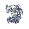
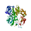
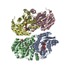
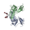
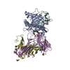
Yeast DNA polymerase alpha, Selenomethionine protein
Components
DNA POLYMERASE ALPHA CATALYTIC SUBUNIT A
Keywords
TRANSFERASE / DNA POLYMERASE / DNA REPLICATION
Function / homology
Function and homology information
Inhibition of replication initiation of damaged DNA by RB1/E2F1 / H3-H4 histone complex chaperone activity / DNA replication initiation / Processive synthesis on the lagging strand / RNA-templated DNA biosynthetic process / Removal of the Flap Intermediate / Polymerase switching / premeiotic DNA replication / alpha DNA polymerase:primase complex / Activation of the pre-replicative complex ...Inhibition of replication initiation of damaged DNA by RB1/E2F1 / H3-H4 histone complex chaperone activity / DNA replication initiation / Processive synthesis on the lagging strand / RNA-templated DNA biosynthetic process / Removal of the Flap Intermediate / Polymerase switching / premeiotic DNA replication / alpha DNA polymerase:primase complex / Activation of the pre-replicative complex / lagging strand elongation / mitotic DNA replication initiation / leading strand elongation / DNA synthesis involved in DNA repair / DNA replication origin binding / DNA replication initiation / replication fork / double-strand break repair / single-stranded DNA binding / 4 iron, 4 sulfur cluster binding / DNA-directed DNA polymerase / DNA-directed DNA polymerase activity / DNA replication / nucleotide binding / chromatin binding / mitochondrion / zinc ion binding Similarity search - Function
B family DNA polymerase, thumb domain, alpha/beta motif / B family DNA polymerase, thumb domain, four helix bundle / Helicase, Ruva Protein; domain 3 - #100 / OB fold (Dihydrolipoamide Acetyltransferase, E2P) - #730 / Alpha-Beta Plaits - #2820 / Helicase, Ruva Protein; domain 3 / DNA polymerase alpha catalytic subunit, N-terminal domain / DNA polymerase alpha, zinc finger domain superfamily / DNA Polymerase alpha zinc finger / DNA polymerase alpha subunit p180 N terminal ...B family DNA polymerase, thumb domain, alpha/beta motif / B family DNA polymerase, thumb domain, four helix bundle / Helicase, Ruva Protein; domain 3 - #100 / OB fold (Dihydrolipoamide Acetyltransferase, E2P) - #730 / Alpha-Beta Plaits - #2820 / Helicase, Ruva Protein; domain 3 / DNA polymerase alpha catalytic subunit, N-terminal domain / DNA polymerase alpha, zinc finger domain superfamily / DNA Polymerase alpha zinc finger / DNA polymerase alpha subunit p180 N terminal / Zinc finger, DNA-directed DNA polymerase, family B, alpha / DNA polymerase alpha catalytic subunit, catalytic domain / Ribonuclease H-like motif / B family DNA polymerase, finger domain / Palm domain of DNA polymerase / B family DNA polymerase, palm domain / Monooxygenase / DNA polymerase family B, thumb domain / DNA-directed DNA polymerase, family B, multifunctional domain / DNA-directed DNA polymerase, family B, conserved site / DNA polymerase family B signature. / DNA polymerase family B / DNA polymerase family B, exonuclease domain / DNA-directed DNA polymerase, family B, exonuclease domain / DNA polymerase, palm domain superfamily / DNA polymerase type-B family / DNA-directed DNA polymerase, family B / Helix non-globular / Ribonuclease H-like superfamily/Ribonuclease H / Special / OB fold (Dihydrolipoamide Acetyltransferase, E2P) / Nucleotidyltransferase; domain 5 / Helix Hairpins / Ribonuclease H superfamily / Ribonuclease H-like superfamily / Alpha-Beta Plaits / DNA/RNA polymerase superfamily / Alpha-Beta Complex / Up-down Bundle / Beta Barrel / 2-Layer Sandwich / Orthogonal Bundle / 3-Layer(aba) Sandwich / Mainly Beta / Mainly Alpha / Alpha Beta Similarity search - Domain/homology
DNAPOLYMERASEALPHACATALYTICSUBUNITA / DNA POLYMERASE I SUBUNIT A / DNA POLYMERASE ALPHA\:PRIMASE COMPLEX P180 SUBUNIT / DNA POLYMERASE- ...DNA POLYMERASE I SUBUNIT A / DNA POLYMERASE ALPHA\:PRIMASE COMPLEX P180 SUBUNIT / DNA POLYMERASE-PRIMASE COMPLEX P180 SUBUNIT / POL ALPHA-PRIMASE COMPLEX P180 SUBUNIT
Mass: 104844.914 Da / Num. of mol.: 1 / Fragment: POLYMERASE DOMAIN, RESIDUES 349-1258 Source method: isolated from a genetically manipulated source Details: SELENOMETHIONINE PROTEIN Source: (gene. exp.) SACCHAROMYCES CEREVISIAE (brewer's yeast) Production host: ESCHERICHIA COLI (E. coli) / Strain (production host): BL21(DE3) / Variant (production host): ROSETTA2 / References: UniProt: P13382, DNA-directed DNA polymerase
Protocol: SINGLE WAVELENGTH / Monochromatic (M) / Laue (L): M / Scattering type: x-ray
Radiation wavelength
Wavelength: 0.9793 Å / Relative weight: 1
Reflection
Resolution: 2.67→73.97 Å / Num. obs: 39626 / % possible obs: 100 % / Observed criterion σ(I): 0 / Redundancy: 19.7 % / Biso Wilson estimate: 45.86 Å2 / Rmerge(I) obs: 0.13 / Net I/σ(I): 17.9
Reflection shell
Resolution: 2.67→2.81 Å / Redundancy: 17.2 % / Rmerge(I) obs: 0.66 / Mean I/σ(I) obs: 4.6 / % possible all: 99.8
-
Processing
Software
Name
Version
Classification
PHENIX
(PHENIX.REFINE: DEV_1078)
refinement
MOSFLM
datareduction
SCALA
datascaling
PHENIX
phasing
Refinement
Method to determine structure: SAD Starting model: NONE Resolution: 2.67→65.8 Å / SU ML: 0.28 / σ(F): 1.35 / Phase error: 24.75 / Stereochemistry target values: ML Details: FOR REFINEMENT, REFMAC, BUSTER AND PHENIX WERE USED. RESIDUES 677-679, 816-847, 1056-1062 1176-1185 AND 1129-1258 ARE DISORDERED AND NOT INCLUDED IN THE STRUCTURE. THE STRUCTURE WAS REFINED ...Details: FOR REFINEMENT, REFMAC, BUSTER AND PHENIX WERE USED. RESIDUES 677-679, 816-847, 1056-1062 1176-1185 AND 1129-1258 ARE DISORDERED AND NOT INCLUDED IN THE STRUCTURE. THE STRUCTURE WAS REFINED WITH RIDING HYDROGEN THE HYDROGENS OF THE LAST REFINEMENT RUN ARE INCLUDED.
Rfactor
Num. reflection
% reflection
Rfree
0.234
1986
5 %
Rwork
0.1952
-
-
obs
0.1972
39564
99.86 %
Solvent computation
Shrinkage radii: 0.9 Å / VDW probe radii: 1.11 Å / Solvent model: FLAT BULK SOLVENT MODEL
Displacement parameters
Biso mean: 56.5 Å2
Refinement step
Cycle: LAST / Resolution: 2.67→65.8 Å
Protein
Nucleic acid
Ligand
Solvent
Total
Num. atoms
6615
0
0
110
6725
Refine LS restraints
Refine-ID
Type
Dev ideal
Number
X-RAY DIFFRACTION
f_bond_d
0.002
6760
X-RAY DIFFRACTION
f_angle_d
0.573
9143
X-RAY DIFFRACTION
f_dihedral_angle_d
12.311
2607
X-RAY DIFFRACTION
f_chiral_restr
0.036
1041
X-RAY DIFFRACTION
f_plane_restr
0.002
1181
LS refinement shell
Resolution (Å)
Rfactor Rfree
Num. reflection Rfree
Rfactor Rwork
Num. reflection Rwork
Refine-ID
% reflection obs (%)
2.6663-2.7329
0.2922
155
0.2429
2587
X-RAY DIFFRACTION
99
2.7329-2.8068
0.2942
139
0.2203
2622
X-RAY DIFFRACTION
100
2.8068-2.8894
0.306
149
0.2208
2657
X-RAY DIFFRACTION
100
2.8894-2.9827
0.2781
117
0.2142
2674
X-RAY DIFFRACTION
100
2.9827-3.0893
0.274
132
0.2158
2662
X-RAY DIFFRACTION
100
3.0893-3.213
0.2915
156
0.2176
2655
X-RAY DIFFRACTION
100
3.213-3.3592
0.2964
124
0.2204
2685
X-RAY DIFFRACTION
100
3.3592-3.5363
0.2644
118
0.2036
2686
X-RAY DIFFRACTION
100
3.5363-3.7578
0.2637
142
0.1926
2664
X-RAY DIFFRACTION
100
3.7578-4.0479
0.1868
139
0.1852
2687
X-RAY DIFFRACTION
100
4.0479-4.4552
0.1961
158
0.1622
2674
X-RAY DIFFRACTION
100
4.4552-5.0997
0.1831
141
0.1529
2716
X-RAY DIFFRACTION
100
5.0997-6.4242
0.2251
137
0.2009
2772
X-RAY DIFFRACTION
100
6.4242-65.8234
0.2127
179
0.2056
2837
X-RAY DIFFRACTION
100
+
About Yorodumi
-
News
-
Feb 9, 2022. New format data for meta-information of EMDB entries
New format data for meta-information of EMDB entries
Version 3 of the EMDB header file is now the official format.
The previous official version 1.9 will be removed from the archive.
In the structure databanks used in Yorodumi, some data are registered as the other names, "COVID-19 virus" and "2019-nCoV". Here are the details of the virus and the list of structure data.
Jan 31, 2019. EMDB accession codes are about to change! (news from PDBe EMDB page)
EMDB accession codes are about to change! (news from PDBe EMDB page)
The allocation of 4 digits for EMDB accession codes will soon come to an end. Whilst these codes will remain in use, new EMDB accession codes will include an additional digit and will expand incrementally as the available range of codes is exhausted. The current 4-digit format prefixed with “EMD-” (i.e. EMD-XXXX) will advance to a 5-digit format (i.e. EMD-XXXXX), and so on. It is currently estimated that the 4-digit codes will be depleted around Spring 2019, at which point the 5-digit format will come into force.
The EM Navigator/Yorodumi systems omit the EMD- prefix.
Related info.:Q: What is EMD? / ID/Accession-code notation in Yorodumi/EM Navigator
Yorodumi is a browser for structure data from EMDB, PDB, SASBDB, etc.
This page is also the successor to EM Navigator detail page, and also detail information page/front-end page for Omokage search.
The word "yorodu" (or yorozu) is an old Japanese word meaning "ten thousand". "mi" (miru) is to see.
Related info.:EMDB / PDB / SASBDB / Comparison of 3 databanks / Yorodumi Search / Aug 31, 2016. New EM Navigator & Yorodumi / Yorodumi Papers / Jmol/JSmol / Function and homology information / Changes in new EM Navigator and Yorodumi
 Movie
Movie Controller


 Open data
 Basic information
 Components
 Keywords
 Function and homology information
 X-RAY DIFFRACTION /
  Authors
 Citation
 Structure visualization
 Downloads & links
 Other downloads


 PDBj
PDBj


 Assembly

 Mass: 18.015 Da / Num. of mol.: 110 / Source method: isolated from a natural source / Formula: H2O
 Sample preparation
 / Beamline: ID23-1 / Wavelength: 0.9793
  Processing