 Movie
Movie Controller

[English] 日本語
 Yorodumi
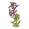
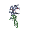
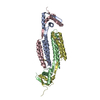
- PDB-4fmn: Structure of the C-terminal domain of the Saccharomyces cerevisia... -
+ Open data

- Basic information
| Entry | Database: PDB / ID: 4fmn | ||||||
|---|---|---|---|---|---|---|---|
| Title | Structure of the C-terminal domain of the Saccharomyces cerevisiae MUTL alpha (MLH1/PMS1) heterodimer bound to a fragment of NTG2 | ||||||
 Components Components |
| ||||||
 Keywords Keywords | HYDROLASE / Mismatch repair / MUTL / endonuclease / Zn-binding protein / DNA damage / DNA repair | ||||||
| Function / homology |  Function and homology information Function and homology informationCleavage of the damaged pyrimidine / meiotic heteroduplex formation / MutLbeta complex / MutLgamma complex / MutLalpha complex / oxidized pyrimidine nucleobase lesion DNA N-glycosylase activity / Mismatch repair (MMR) directed by MSH2:MSH6 (MutSalpha) / meiotic mismatch repair / base-excision repair, AP site formation / mismatched DNA binding ...Cleavage of the damaged pyrimidine / meiotic heteroduplex formation / MutLbeta complex / MutLgamma complex / MutLalpha complex / oxidized pyrimidine nucleobase lesion DNA N-glycosylase activity / Mismatch repair (MMR) directed by MSH2:MSH6 (MutSalpha) / meiotic mismatch repair / base-excision repair, AP site formation / mismatched DNA binding / reciprocal meiotic recombination / Hydrolases; Glycosylases; Hydrolysing N-glycosyl compounds / ATP-dependent DNA damage sensor activity / mismatch repair / DNA-(apurinic or apyrimidinic site) endonuclease activity / class I DNA-(apurinic or apyrimidinic site) endonuclease activity / DNA-(apurinic or apyrimidinic site) lyase / nucleotide-excision repair / base-excision repair / 4 iron, 4 sulfur cluster binding / ATP hydrolysis activity / mitochondrion / DNA binding / ATP binding / metal ion binding / nucleus / cytoplasm Similarity search - Function | ||||||
| Biological species |  | ||||||
| Method |  X-RAY DIFFRACTION / SYNCHROTRON / MOLECULAR REPLACEMENT / Resolution: 2.69 Å X-RAY DIFFRACTION / SYNCHROTRON / MOLECULAR REPLACEMENT / Resolution: 2.69 Å | ||||||
 Authors Authors | Gueneau, E. / Legrand, P. / Charbonnier, J.B. | ||||||
 Citation Citation | Journal: Nat.Struct.Mol.Biol. / Year: 2013 Title: Structure of the MutL alpha C-terminal domain reveals how Mlh1 contributes to Pms1 endonuclease site. Authors: Gueneau, E. / Dherin, C. / Legrand, P. / Tellier-Lebegue, C. / Gilquin, B. / Bonnesoeur, P. / Londino, F. / Quemener, C. / Le Du, M.H. / Marquez, J.A. / Moutiez, M. / Gondry, M. / Boiteux, S. / Charbonnier, J.B. | ||||||
| History |
|
- Structure visualization
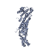
| Structure viewer | Molecule: MolmilJmol/JSmol |
|---|
- Downloads & links
-Download
| PDBx/mmCIF format | 4fmn.cif.gz | 120.9 KB | Display | PDBx/mmCIF format |
|---|---|---|---|---|
| PDB format | pdb4fmn.ent.gz | 91.2 KB | Display | PDB format |
| PDBx/mmJSON format | 4fmn.json.gz | Tree view | PDBx/mmJSON format | |
| Others |  Other downloads Other downloads |
-Validation report
| Arichive directory | https://data.pdbj.org/pub/pdb/validation_reports/fm/4fmnftp://data.pdbj.org/pub/pdb/validation_reports/fm/4fmn | HTTPS FTP |
|---|
-Related structure data
| Related structure data |  4e4wSC  4fmoC C: citing same article ( S: Starting model for refinement |
|---|---|
| Similar structure data |


-Links
 PDBj
PDBj

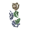
- Assembly
| Deposited unit | 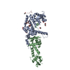
| ||||||||
|---|---|---|---|---|---|---|---|---|---|
| 1 |
| ||||||||
| Unit cell |
|
-Components
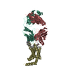
-DNA mismatch repair protein ... , 2 types, 2 molecules AB
| #1: Protein | Mass: 33239.102 Da / Num. of mol.: 1 / Fragment: UNP residues 483-769 Source method: isolated from a genetically manipulated source Source: (gene. exp.) Strain: ATCC 204508 / S288c / Gene: MLH1, PMS2, YMR167W, YM8520.16 / Production host:  |
|---|---|
| #2: Protein | Mass: 27948.195 Da / Num. of mol.: 1 / Fragment: UNP residues 635-873 Source method: isolated from a genetically manipulated source Source: (gene. exp.) Strain: ATCC 204508 / S288c / Gene: PMS1, YNL082W, N2317 / Production host: |
-Protein/peptide , 1 types, 1 molecules C
| #3: Protein/peptide | Mass: 1085.342 Da / Num. of mol.: 1 / Fragment: UNP residues 22-29 / Source method: obtained synthetically / Details: Synthetic peptide / Source: (synth.) |
|---|
-Non-polymers , 4 types, 144 molecules 


| #4: Chemical | ChemComp-GOL /  Mass: 92.094 Da / Num. of mol.: 5 / Source method: obtained synthetically / Formula: C3H8O3 Mass: 92.094 Da / Num. of mol.: 5 / Source method: obtained synthetically / Formula: C3H8O3#5: Chemical | ChemComp-EDO /  Mass: 62.068 Da / Num. of mol.: 6 / Source method: obtained synthetically / Formula: C2H6O2 Mass: 62.068 Da / Num. of mol.: 6 / Source method: obtained synthetically / Formula: C2H6O2#6: Chemical |  Mass: 65.409 Da / Num. of mol.: 2 / Source method: obtained synthetically / Formula: Zn Mass: 65.409 Da / Num. of mol.: 2 / Source method: obtained synthetically / Formula: Zn#7: Water | ChemComp-HOH / | Mass: 18.015 Da / Num. of mol.: 131 / Source method: isolated from a natural source / Formula: H2O |
|---|
-Details
| Has protein modification | Y |
|---|
-Experimental details
-Experiment
| Experiment | Method: X-RAY DIFFRACTION / Number of used crystals: 1 |
|---|
- Sample preparation
| Crystal | Density Matthews: 3.77 Å3/Da / Density % sol: 67.34 % |
|---|---|
| Crystal grow | Temperature: 293 K / Method: vapor diffusion, sitting drop / pH: 7 Details: PEG, pH 7, VAPOR DIFFUSION, SITTING DROP, temperature 293K |
-Data collection
| Diffraction | Mean temperature: 100 K |
|---|---|
| Diffraction source | Source: SYNCHROTRON / Site: SOLEIL  / Beamline: PROXIMA 1 / Wavelength: 0.9801 Å / Beamline: PROXIMA 1 / Wavelength: 0.9801 Å |
| Detector | Type: ADSC QUANTUM 315r / Detector: CCD / Date: Sep 24, 2011 |
| Radiation | Protocol: SINGLE WAVELENGTH / Monochromatic (M) / Laue (L): M / Scattering type: x-ray |
| Radiation wavelength | Wavelength: 0.9801 Å / Relative weight: 1 |
| Reflection | Resolution: 2.69→50 Å / Num. all: 25931 / Num. obs: 25750 / % possible obs: 99.3 % / Redundancy: 5.6 % / Biso Wilson estimate: 66.07 Å2 / Rsym value: 0.098 |
| Reflection shell | Resolution: 2.69→2.86 Å / Redundancy: 5.6 % / Mean I/σ(I) obs: 2.3 / Rsym value: 0.768 / % possible all: 96.8 |
- Processing
| Software |
| ||||||||||||||||||||||||||||||||||||||||||||||||||||||||||||||||||||||||||||||||||||||||||||||||||||||||||||||||||
|---|---|---|---|---|---|---|---|---|---|---|---|---|---|---|---|---|---|---|---|---|---|---|---|---|---|---|---|---|---|---|---|---|---|---|---|---|---|---|---|---|---|---|---|---|---|---|---|---|---|---|---|---|---|---|---|---|---|---|---|---|---|---|---|---|---|---|---|---|---|---|---|---|---|---|---|---|---|---|---|---|---|---|---|---|---|---|---|---|---|---|---|---|---|---|---|---|---|---|---|---|---|---|---|---|---|---|---|---|---|---|---|---|---|---|---|
| Refinement | Method to determine structure: MOLECULAR REPLACEMENT Starting model: 4E4W Resolution: 2.69→31.63 Å / Cor.coef. Fo:Fc: 0.9421 / Cor.coef. Fo:Fc free: 0.9315 / SU R Cruickshank DPI: 0.304 / Cross valid method: THROUGHOUT / σ(F): 0
| ||||||||||||||||||||||||||||||||||||||||||||||||||||||||||||||||||||||||||||||||||||||||||||||||||||||||||||||||||
| Displacement parameters | Biso mean: 85.23 Å2
| ||||||||||||||||||||||||||||||||||||||||||||||||||||||||||||||||||||||||||||||||||||||||||||||||||||||||||||||||||
| Refine analyze | Luzzati coordinate error obs: 0.398 Å | ||||||||||||||||||||||||||||||||||||||||||||||||||||||||||||||||||||||||||||||||||||||||||||||||||||||||||||||||||
| Refinement step | Cycle: LAST / Resolution: 2.69→31.63 Å
| ||||||||||||||||||||||||||||||||||||||||||||||||||||||||||||||||||||||||||||||||||||||||||||||||||||||||||||||||||
| Refine LS restraints |
| ||||||||||||||||||||||||||||||||||||||||||||||||||||||||||||||||||||||||||||||||||||||||||||||||||||||||||||||||||
| LS refinement shell | Resolution: 2.69→2.8 Å / Total num. of bins used: 13
| ||||||||||||||||||||||||||||||||||||||||||||||||||||||||||||||||||||||||||||||||||||||||||||||||||||||||||||||||||
| Refinement TLS params. | Method: refined / Refine-ID: X-RAY DIFFRACTION
| ||||||||||||||||||||||||||||||||||||||||||||||||||||||||||||||||||||||||||||||||||||||||||||||||||||||||||||||||||
| Refinement TLS group |
|
