 Movie
Movie Controller

[English] 日本語
 Yorodumi
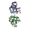
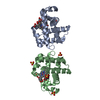
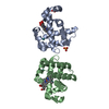
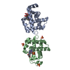
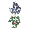
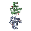
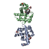
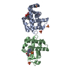
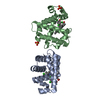
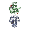
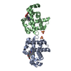
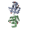
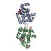
- PDB-4fh6: Structure of DHP A in complex with 2,4,6-tribromophenol in 10% DMSO -
+ Open data

- Basic information
| Entry | Database: PDB / ID: 4fh6 | ||||||
|---|---|---|---|---|---|---|---|
| Title | Structure of DHP A in complex with 2,4,6-tribromophenol in 10% DMSO | ||||||
 Components Components | Dehaloperoxidase A | ||||||
 Keywords Keywords | OXIDOREDUCTASE / peroxidase / globin | ||||||
| Function / homology |  Function and homology information Function and homology informationoxygen carrier activity / peroxidase activity / oxygen binding / heme binding / metal ion binding Similarity search - Function | ||||||
| Biological species |   Amphitrite ornata (invertebrata) Amphitrite ornata (invertebrata) | ||||||
| Method |  X-RAY DIFFRACTION / SYNCHROTRON / MOLECULAR REPLACEMENT / Resolution: 1.44 Å X-RAY DIFFRACTION / SYNCHROTRON / MOLECULAR REPLACEMENT / Resolution: 1.44 Å | ||||||
 Authors Authors | de Serrano, V.S. / Franzen, S. | ||||||
 Citation Citation | Journal: Biochemistry / Year: 2013 Title: Structural and Kinetic Study of an Internal Substrate Binding Site in Dehaloperoxidase-Hemoglobin A from Amphitrite ornata. Authors: Zhao, J. / de Serrano, V. / Zhao, J. / Le, P. / Franzen, S. | ||||||
| History |
|
- Structure visualization
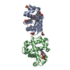
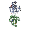
| Structure viewer | Molecule: MolmilJmol/JSmol |
|---|
- Downloads & links
-Download
| PDBx/mmCIF format | 4fh6.cif.gz | 166.6 KB | Display | PDBx/mmCIF format |
|---|---|---|---|---|
| PDB format | pdb4fh6.ent.gz | 134.1 KB | Display | PDB format |
| PDBx/mmJSON format | 4fh6.json.gz | Tree view | PDBx/mmJSON format | |
| Others |  Other downloads Other downloads |
-Validation report
| Arichive directory | https://data.pdbj.org/pub/pdb/validation_reports/fh/4fh6ftp://data.pdbj.org/pub/pdb/validation_reports/fh/4fh6 | HTTPS FTP |
|---|
-Related structure data
| Related structure data |  4fh7C  4ilzC 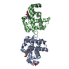 2qfkS C: citing same article ( S: Starting model for refinement |
|---|---|
| Similar structure data |


-Links
 PDBj
PDBj


- Assembly
| Deposited unit | 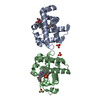
| ||||||||
|---|---|---|---|---|---|---|---|---|---|
| 1 |
| ||||||||
| Unit cell |
|
-Components
-Protein , 1 types, 2 molecules AB
| #1: Protein | Mass: 15548.597 Da / Num. of mol.: 2 Source method: isolated from a genetically manipulated source Source: (gene. exp.) Amphitrite ornata (invertebrata) / Gene: dhpA / Plasmid: pET16b / Production host:  |
|---|
-Non-polymers , 6 types, 248 molecules 


| #2: Chemical |  Mass: 616.487 Da / Num. of mol.: 2 / Source method: obtained synthetically / Formula: C34H32FeN4O4 Mass: 616.487 Da / Num. of mol.: 2 / Source method: obtained synthetically / Formula: C34H32FeN4O4#3: Chemical |  Mass: 96.063 Da / Num. of mol.: 3 / Source method: obtained synthetically / Formula: SO4 Mass: 96.063 Da / Num. of mol.: 3 / Source method: obtained synthetically / Formula: SO4#4: Chemical |  Mass: 330.799 Da / Num. of mol.: 2 / Source method: obtained synthetically / Formula: C6H3Br3O Mass: 330.799 Da / Num. of mol.: 2 / Source method: obtained synthetically / Formula: C6H3Br3O#5: Chemical |  Mass: 31.999 Da / Num. of mol.: 2 / Source method: obtained synthetically / Formula: O2 Mass: 31.999 Da / Num. of mol.: 2 / Source method: obtained synthetically / Formula: O2#6: Chemical | ChemComp-DMS / |  Mass: 78.133 Da / Num. of mol.: 1 / Source method: obtained synthetically / Formula: C2H6OS / Comment: DMSO, precipitant*YM Mass: 78.133 Da / Num. of mol.: 1 / Source method: obtained synthetically / Formula: C2H6OS / Comment: DMSO, precipitant*YM#7: Water | ChemComp-HOH / | Mass: 18.015 Da / Num. of mol.: 238 / Source method: isolated from a natural source / Formula: H2O |
|---|
-Experimental details
-Experiment
| Experiment | Method: X-RAY DIFFRACTION / Number of used crystals: 1 |
|---|
- Sample preparation
| Crystal | Density Matthews: 2.15 Å3/Da / Density % sol: 42.79 % |
|---|---|
| Crystal grow | Temperature: 277 K / Method: vapor diffusion, hanging drop / pH: 5.9 Details: 0.2 M Ammonium Sulfate, 32% w/v PEG 4000, pH 5.9, VAPOR DIFFUSION, HANGING DROP, temperature 277K |
-Data collection
| Diffraction | Mean temperature: 100 K |
|---|---|
| Diffraction source | Source: SYNCHROTRON / Site: APS  / Beamline: 22-BM / Wavelength: 0.91339 Å / Beamline: 22-BM / Wavelength: 0.91339 Å |
| Detector | Type: MARMOSAIC 225 mm CCD / Detector: CCD / Date: Mar 14, 2010 Details: Rosenbaum-Rock double-crystal monochromator: sagitally focusing 2nd crystal, Rosenbaum-Rock vertical focusing mirror |
| Radiation | Monochromator: double crystal / Protocol: SINGLE WAVELENGTH / Monochromatic (M) / Laue (L): M / Scattering type: x-ray |
| Radiation wavelength | Wavelength: 0.91339 Å / Relative weight: 1 |
| Reflection | Resolution: 1.44→35 Å / Num. all: 46777 / Num. obs: 46300 / % possible obs: 98.98 % / Observed criterion σ(I): 2 / Redundancy: 3.6 % / Biso Wilson estimate: 8.2 Å2 / Rmerge(I) obs: 0.089 / Net I/σ(I): 11.9 |
| Reflection shell | Resolution: 1.44→1.447 Å / Redundancy: 3.5 % / Rmerge(I) obs: 0.413 / Mean I/σ(I) obs: 2.69 / Num. unique all: 3423 / % possible all: 98.61 |
- Processing
| Software |
| |||||||||||||||||||||||||||||||||||||||||||||||||||||||||||||||||||||||||||
|---|---|---|---|---|---|---|---|---|---|---|---|---|---|---|---|---|---|---|---|---|---|---|---|---|---|---|---|---|---|---|---|---|---|---|---|---|---|---|---|---|---|---|---|---|---|---|---|---|---|---|---|---|---|---|---|---|---|---|---|---|---|---|---|---|---|---|---|---|---|---|---|---|---|---|---|---|
| Refinement | Method to determine structure: MOLECULAR REPLACEMENT Starting model: PDB ENTRY 2QFK Resolution: 1.44→35 Å / Cor.coef. Fo:Fc: 0.951 / Cor.coef. Fo:Fc free: 0.928 / SU B: 2.357 / SU ML: 0.041 / Isotropic thermal model: Anisotropic / Cross valid method: THROUGHOUT / σ(I): 2 / ESU R: 0.028 / ESU R Free: 0.019 / Stereochemistry target values: MAXIMUM LIKELIHOOD Details: HYDROGENS HAVE BEEN ADDED IN THE RIDING POSITIONS. The ligand TBP was positioned in the structure by making use of anomalous scattering of bromine atoms to the anomalous difference maps in ...Details: HYDROGENS HAVE BEEN ADDED IN THE RIDING POSITIONS. The ligand TBP was positioned in the structure by making use of anomalous scattering of bromine atoms to the anomalous difference maps in order to locate and position bromine atoms of the tribromophenol molecule
| |||||||||||||||||||||||||||||||||||||||||||||||||||||||||||||||||||||||||||
| Solvent computation | Ion probe radii: 0.8 Å / Shrinkage radii: 0.8 Å / VDW probe radii: 1.2 Å / Solvent model: MASK | |||||||||||||||||||||||||||||||||||||||||||||||||||||||||||||||||||||||||||
| Displacement parameters | Biso mean: 8.181 Å2
| |||||||||||||||||||||||||||||||||||||||||||||||||||||||||||||||||||||||||||
| Refinement step | Cycle: LAST / Resolution: 1.44→35 Å
| |||||||||||||||||||||||||||||||||||||||||||||||||||||||||||||||||||||||||||
| Refine LS restraints |
| |||||||||||||||||||||||||||||||||||||||||||||||||||||||||||||||||||||||||||
| LS refinement shell | Resolution: 1.44→1.477 Å / Total num. of bins used: 20
|
