 Movie
Movie Controller

[English] 日本語
 Yorodumi
- PDB-1ew6: THE CRYSTAL STRUCTURE AND AMINO ACID SEQUENCE OF DEHALOPEROXIDASE... -
+ Open data

- Basic information
| Entry | Database: PDB / ID: 1ew6 | ||||||
|---|---|---|---|---|---|---|---|
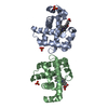
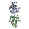
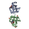
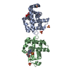
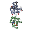
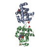
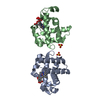
| Title | THE CRYSTAL STRUCTURE AND AMINO ACID SEQUENCE OF DEHALOPEROXIDASE FROM AMPHITRITE ORNATA INDICATE COMMON ANCESTRY WITH GLOBINS | ||||||
 Components Components | DEHALOPEROXIDASE | ||||||
 Keywords Keywords | OXIDOREDUCTASE / GLOBIN / HALOPHENOLS | ||||||
| Function / homology |  Function and homology information Function and homology informationoxygen carrier activity / peroxidase activity / oxygen binding / heme binding / metal ion binding Similarity search - Function | ||||||
| Biological species |   Amphitrite ornata (invertebrata) Amphitrite ornata (invertebrata) | ||||||
| Method |  X-RAY DIFFRACTION / Resolution: 1.78 Å X-RAY DIFFRACTION / Resolution: 1.78 Å | ||||||
 Authors Authors | Lebioda, L. | ||||||
 Citation Citation | Journal: J.Biol.Chem. / Year: 2000 Title: The crystal structure and amino acid sequence of dehaloperoxidase from Amphitrite ornata indicate common ancestry with globins. Authors: LaCount, M.W. / Zhang, E. / Chen, Y.P. / Han, K. / Whitton, M.M. / Lincoln, D.E. / Woodin, S.A. / Lebioda, L. | ||||||
| History |
|
- Structure visualization
| Structure viewer | Molecule: MolmilJmol/JSmol |
|---|
- Downloads & links
-Download
| PDBx/mmCIF format | 1ew6.cif.gz | 70.2 KB | Display | PDBx/mmCIF format |
|---|---|---|---|---|
| PDB format | pdb1ew6.ent.gz | 53.1 KB | Display | PDB format |
| PDBx/mmJSON format | 1ew6.json.gz | Tree view | PDBx/mmJSON format | |
| Others |  Other downloads Other downloads |
-Validation report
| Arichive directory | https://data.pdbj.org/pub/pdb/validation_reports/ew/1ew6ftp://data.pdbj.org/pub/pdb/validation_reports/ew/1ew6 | HTTPS FTP |
|---|
-Related structure data


-Links
 PDBj
PDBj


- Assembly
| Deposited unit | 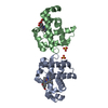
| ||||||||
|---|---|---|---|---|---|---|---|---|---|
| 1 |
| ||||||||
| Unit cell |
|
-Components
| #1: Protein | Mass: 15548.597 Da / Num. of mol.: 2 / Source method: isolated from a natural source / Details: POLYCHAETE / Source: (natural) Amphitrite ornata (invertebrata) / References: UniProt: Q9NAV8#2: Chemical |   Mass: 96.063 Da / Num. of mol.: 2 / Source method: obtained synthetically / Formula: SO4 Mass: 96.063 Da / Num. of mol.: 2 / Source method: obtained synthetically / Formula: SO4#3: Chemical |   Mass: 616.487 Da / Num. of mol.: 2 / Source method: obtained synthetically / Formula: C34H32FeN4O4 Mass: 616.487 Da / Num. of mol.: 2 / Source method: obtained synthetically / Formula: C34H32FeN4O4#4: Water | ChemComp-HOH / |  Mass: 18.015 Da / Num. of mol.: 107 / Source method: isolated from a natural source / Formula: H2O Mass: 18.015 Da / Num. of mol.: 107 / Source method: isolated from a natural source / Formula: H2O |
|---|
-Experimental details
-Experiment
| Experiment | Method: X-RAY DIFFRACTION / Number of used crystals: 1 |
|---|
- Sample preparation
| Crystal | Density Matthews: 2.3 Å3/Da / Density % sol: 46.46 % | ||||||||||||||||||||
|---|---|---|---|---|---|---|---|---|---|---|---|---|---|---|---|---|---|---|---|---|---|
| Crystal grow | Temperature: 298 K / Method: vapor diffusion, hanging drop / pH: 7 Details: PEG 8000, 200 MM AMMONIUM SULFATE, pH 7.0, VAPOR DIFFUSION, HANGING DROP, temperature 298K | ||||||||||||||||||||
| Crystal grow | *PLUS Temperature: 277 K / pH: 6.5 / Details: Zhang, E., (1996) Acta Crystallogr., D52, 1191. | ||||||||||||||||||||
| Components of the solutions | *PLUS
|
-Data collection
| Diffraction | Mean temperature: 298 K |
|---|---|
| Diffraction source | Source: ROTATING ANODE / Type: RIGAKU RU200 / Wavelength: 1.5418 |
| Detector | Type: RIGAKU RAXIS II / Detector: IMAGE PLATE / Date: Oct 12, 1995 |
| Radiation | Protocol: SINGLE WAVELENGTH / Monochromatic (M) / Laue (L): M / Scattering type: x-ray |
| Radiation wavelength | Wavelength: 1.5418 Å / Relative weight: 1 |
| Reflection | Resolution: 1.78→10 Å / Num. obs: 24637 / Observed criterion σ(F): 3 / Observed criterion σ(I): 1 / Redundancy: 2.3 % / Rmerge(I) obs: 0.051 |
| Reflection | *PLUS Num. measured all: 66563 |
- Processing
| Software |
| ||||||||||||||||||||
|---|---|---|---|---|---|---|---|---|---|---|---|---|---|---|---|---|---|---|---|---|---|
| Refinement | Resolution: 1.78→8 Å / σ(F): 3 / σ(I): 1 / Stereochemistry target values: ENGH & HUBER
| ||||||||||||||||||||
| Refinement step | Cycle: LAST / Resolution: 1.78→8 Å
| ||||||||||||||||||||
| Refine LS restraints |
| ||||||||||||||||||||
| Software | *PLUS Name: CNS / Classification: refinement | ||||||||||||||||||||
| Refinement | *PLUS Lowest resolution: 8 Å / σ(F): 3 / Rfactor obs: 0.197 | ||||||||||||||||||||
| Solvent computation | *PLUS | ||||||||||||||||||||
| Displacement parameters | *PLUS | ||||||||||||||||||||
| Refine LS restraints | *PLUS
|
