 Movie
Movie Controller

[English] 日本語
 Yorodumi
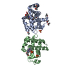
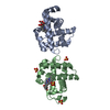
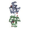
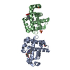
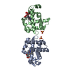
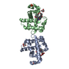
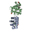
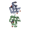
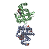
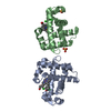
- PDB-4fh7: Structure of DHP A in complex with 2,4,6-tribromophenol in 20% me... -
+ Open data

- Basic information
| Entry | Database: PDB / ID: 4fh7 | ||||||
|---|---|---|---|---|---|---|---|
| Title | Structure of DHP A in complex with 2,4,6-tribromophenol in 20% methanol | ||||||
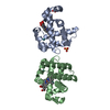
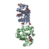
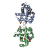
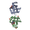
 Components Components | Dehaloperoxidase A | ||||||
 Keywords Keywords | OXIDOREDUCTASE / peroxidase / globin | ||||||
| Function / homology |  Function and homology information Function and homology informationoxygen carrier activity / peroxidase activity / oxygen binding / heme binding / metal ion binding Similarity search - Function | ||||||
| Biological species |   Amphitrite ornata (invertebrata) Amphitrite ornata (invertebrata) | ||||||
| Method |  X-RAY DIFFRACTION / SYNCHROTRON / MOLECULAR REPLACEMENT / Resolution: 1.74 Å X-RAY DIFFRACTION / SYNCHROTRON / MOLECULAR REPLACEMENT / Resolution: 1.74 Å | ||||||
 Authors Authors | de Serrano, V.S. / Franzen, S. | ||||||
 Citation Citation | Journal: Biochemistry / Year: 2013 Title: Structural and Kinetic Study of an Internal Substrate Binding Site in Dehaloperoxidase-Hemoglobin A from Amphitrite ornata. Authors: Zhao, J. / de Serrano, V. / Zhao, J. / Le, P. / Franzen, S. | ||||||
| History |
|
- Structure visualization
| Structure viewer | Molecule: MolmilJmol/JSmol |
|---|
- Downloads & links
-Download
| PDBx/mmCIF format | 4fh7.cif.gz | 92.9 KB | Display | PDBx/mmCIF format |
|---|---|---|---|---|
| PDB format | pdb4fh7.ent.gz | 72.9 KB | Display | PDB format |
| PDBx/mmJSON format | 4fh7.json.gz | Tree view | PDBx/mmJSON format | |
| Others |  Other downloads Other downloads |
-Validation report
| Arichive directory | https://data.pdbj.org/pub/pdb/validation_reports/fh/4fh7ftp://data.pdbj.org/pub/pdb/validation_reports/fh/4fh7 | HTTPS FTP |
|---|
-Related structure data
| Related structure data |  4fh6C  4ilzC 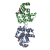 2qfkS C: citing same article ( S: Starting model for refinement |
|---|---|
| Similar structure data |


-Links
 PDBj
PDBj


- Assembly
| Deposited unit | 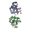
| ||||||||
|---|---|---|---|---|---|---|---|---|---|
| 1 |
| ||||||||
| Unit cell |
|
-Components
-Protein , 1 types, 2 molecules AB
| #1: Protein | Mass: 15548.597 Da / Num. of mol.: 2 Source method: isolated from a genetically manipulated source Source: (gene. exp.) Amphitrite ornata (invertebrata) / Gene: dhpA / Plasmid: pET16b / Production host:  |
|---|
-Non-polymers , 6 types, 259 molecules 


| #2: Chemical |  Mass: 616.487 Da / Num. of mol.: 2 / Source method: obtained synthetically / Formula: C34H32FeN4O4 Mass: 616.487 Da / Num. of mol.: 2 / Source method: obtained synthetically / Formula: C34H32FeN4O4#3: Chemical | ChemComp-TBP / |  Mass: 330.799 Da / Num. of mol.: 1 / Source method: obtained synthetically / Formula: C6H3Br3O Mass: 330.799 Da / Num. of mol.: 1 / Source method: obtained synthetically / Formula: C6H3Br3O#4: Chemical | ChemComp-OXY / |  Mass: 31.999 Da / Num. of mol.: 1 / Source method: obtained synthetically / Formula: O2 Mass: 31.999 Da / Num. of mol.: 1 / Source method: obtained synthetically / Formula: O2#5: Chemical | ChemComp-SO4 /  Mass: 96.063 Da / Num. of mol.: 8 / Source method: obtained synthetically / Formula: SO4 Mass: 96.063 Da / Num. of mol.: 8 / Source method: obtained synthetically / Formula: SO4#6: Chemical | ChemComp-MOH /  Mass: 32.042 Da / Num. of mol.: 4 / Source method: obtained synthetically / Formula: CH4O Mass: 32.042 Da / Num. of mol.: 4 / Source method: obtained synthetically / Formula: CH4O#7: Water | ChemComp-HOH / | Mass: 18.015 Da / Num. of mol.: 243 / Source method: isolated from a natural source / Formula: H2O |
|---|
-Experimental details
-Experiment
| Experiment | Method: X-RAY DIFFRACTION / Number of used crystals: 1 |
|---|
- Sample preparation
| Crystal | Density Matthews: 2.15 Å3/Da / Density % sol: 42.7 % |
|---|---|
| Crystal grow | Temperature: 277 K / Method: vapor diffusion, hanging drop / pH: 5.9 Details: 0.2 M Ammonium Sulfate, 32% w/v PEG 4000, pH 5.9, VAPOR DIFFUSION, HANGING DROP, temperature 277K |
-Data collection
| Diffraction | Mean temperature: 100 K |
|---|---|
| Diffraction source | Source: SYNCHROTRON / Site: APS  / Beamline: 22-BM / Wavelength: 0.91339 Å / Beamline: 22-BM / Wavelength: 0.91339 Å |
| Detector | Type: MARMOSAIC 225 mm CCD / Detector: CCD / Date: Mar 14, 2010 Details: Rosenbaum-Rock double-crystal monochromator, sagitally focusing 2nd crystal, Rosenbaum-Rock vertical focusing mirror |
| Radiation | Monochromator: double crystaL / Protocol: SINGLE WAVELENGTH / Monochromatic (M) / Laue (L): M / Scattering type: x-ray |
| Radiation wavelength | Wavelength: 0.91339 Å / Relative weight: 1 |
| Reflection | Resolution: 1.74→35 Å / Num. all: 26670 / Num. obs: 26593 / % possible obs: 99.71 % / Observed criterion σ(I): 2 / Redundancy: 3.7 % / Biso Wilson estimate: 10.7 Å2 / Rmerge(I) obs: 0.116 / Net I/σ(I): 5.1 |
| Reflection shell | Resolution: 1.74→1.784 Å / Redundancy: 3.9 % / Rmerge(I) obs: 0.428 / Mean I/σ(I) obs: 4 / Num. unique all: 1946 / % possible all: 98.38 |
- Processing
| Software |
| |||||||||||||||||||||||||||||||||||||||||||||
|---|---|---|---|---|---|---|---|---|---|---|---|---|---|---|---|---|---|---|---|---|---|---|---|---|---|---|---|---|---|---|---|---|---|---|---|---|---|---|---|---|---|---|---|---|---|---|
| Refinement | Method to determine structure: MOLECULAR REPLACEMENT Starting model: PDB ENTRY 2QFK Resolution: 1.74→35 Å / Cor.coef. Fo:Fc: 0.931 / Cor.coef. Fo:Fc free: 0.893 / SU B: 2.988 / SU ML: 0.095 / Isotropic thermal model: Isotropic / Cross valid method: THROUGHOUT / σ(I): 2 / ESU R: 0.044 / ESU R Free: 0.037 / Stereochemistry target values: MAXIMUM LIKELIHOOD / Details: HYDROGENS HAVE BEEN USED IF PRESENT IN THE INPUT
| |||||||||||||||||||||||||||||||||||||||||||||
| Solvent computation | Ion probe radii: 0.8 Å / Shrinkage radii: 0.8 Å / VDW probe radii: 1.2 Å / Solvent model: MASK | |||||||||||||||||||||||||||||||||||||||||||||
| Displacement parameters | Biso mean: 9.608 Å2
| |||||||||||||||||||||||||||||||||||||||||||||
| Refinement step | Cycle: LAST / Resolution: 1.74→35 Å
| |||||||||||||||||||||||||||||||||||||||||||||
| Refine LS restraints |
| |||||||||||||||||||||||||||||||||||||||||||||
| LS refinement shell | Resolution: 1.74→1.784 Å / Total num. of bins used: 20
|
