 Movie
Movie Controller

[English] 日本語
 Yorodumi
- PDB-4fdt: Crystal Structure of a Multiple Inositol Polyphosphate Phosphatase -
+ Open data

- Basic information
| Entry | Database: PDB / ID: 4fdt | ||||||
|---|---|---|---|---|---|---|---|
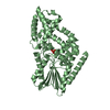
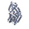
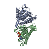
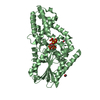
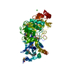
| Title | Crystal Structure of a Multiple Inositol Polyphosphate Phosphatase | ||||||
 Components Components | Putative multiple inositol polyphosphate histidine phosphatase 1 | ||||||
 Keywords Keywords | HYDROLASE / Phosphatase | ||||||
| Function / homology |  Function and homology information Function and homology informationmultiple inositol-polyphosphate phosphatase / 2,3-bisphosphoglycerate 3-phosphatase / bisphosphoglycerate 3-phosphatase activity / membrane Similarity search - Function | ||||||
| Biological species |  Bacteroides thetaiotaomicron (bacteria) Bacteroides thetaiotaomicron (bacteria) | ||||||
| Method |  X-RAY DIFFRACTION / SYNCHROTRON / SAD / Resolution: 1.93 Å X-RAY DIFFRACTION / SYNCHROTRON / SAD / Resolution: 1.93 Å | ||||||
 Authors Authors | Li, A.W.H. / Hemmings, A.M. | ||||||
 Citation Citation | Journal: Cell Rep / Year: 2014 Title: A Bacterial Homolog of a Eukaryotic Inositol Phosphate Signaling Enzyme Mediates Cross-kingdom Dialog in the Mammalian Gut. Authors: Stentz, R. / Osborne, S. / Horn, N. / Li, A.W. / Hautefort, I. / Bongaerts, R. / Rouyer, M. / Bailey, P. / Shears, S.B. / Hemmings, A.M. / Brearley, C.A. / Carding, S.R. | ||||||
| History |
|
- Structure visualization
| Structure viewer | Molecule: MolmilJmol/JSmol |
|---|
- Downloads & links
-Download
| PDBx/mmCIF format | 4fdt.cif.gz | 352.1 KB | Display | PDBx/mmCIF format |
|---|---|---|---|---|
| PDB format | pdb4fdt.ent.gz | 289.2 KB | Display | PDB format |
| PDBx/mmJSON format | 4fdt.json.gz | Tree view | PDBx/mmJSON format | |
| Others |  Other downloads Other downloads |
-Validation report
| Arichive directory | https://data.pdbj.org/pub/pdb/validation_reports/fd/4fdtftp://data.pdbj.org/pub/pdb/validation_reports/fd/4fdt | HTTPS FTP |
|---|
-Related structure data


-Links
 PDBj
PDBj


- Assembly
| Deposited unit | 
| ||||||||
|---|---|---|---|---|---|---|---|---|---|
| 1 | 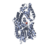
| ||||||||
| 2 | 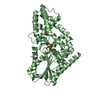
| ||||||||
| Unit cell |
|
-Components
| #1: Protein | Mass: 49203.426 Da / Num. of mol.: 2 / Fragment: UNP residues 21-425 Source method: isolated from a genetically manipulated source Source: (gene. exp.) Bacteroides thetaiotaomicron (bacteria)Strain: ATCC 29148 / DSM 2079 / NCTC 10582 / E50 / VPI-5482 / Gene: BT_4744 / Plasmid: pET15b / Production host: #2: Chemical |   Mass: 94.971 Da / Num. of mol.: 2 / Source method: obtained synthetically / Formula: PO4 Mass: 94.971 Da / Num. of mol.: 2 / Source method: obtained synthetically / Formula: PO4#3: Chemical |   Mass: 62.068 Da / Num. of mol.: 2 / Source method: obtained synthetically / Formula: C2H6O2 Mass: 62.068 Da / Num. of mol.: 2 / Source method: obtained synthetically / Formula: C2H6O2#4: Water | ChemComp-HOH / |  Mass: 18.015 Da / Num. of mol.: 451 / Source method: isolated from a natural source / Formula: H2O Mass: 18.015 Da / Num. of mol.: 451 / Source method: isolated from a natural source / Formula: H2O |
|---|
-Experimental details
-Experiment
| Experiment | Method: X-RAY DIFFRACTION / Number of used crystals: 1 |
|---|
- Sample preparation
| Crystal | Density Matthews: 2.34 Å3/Da / Density % sol: 47.42 % |
|---|---|
| Crystal grow | Temperature: 289 K / Method: vapor diffusion / pH: 5 Details: 0.2M Ammonium acetate, pH5.0, 18% PEG 3350, Protein 1mg/ml, VAPOR DIFFUSION, temperature 289K |
-Data collection
| Diffraction | Mean temperature: 100 K |
|---|---|
| Diffraction source | Source: SYNCHROTRON / Site: Diamond  / Beamline: I24 / Wavelength: 0.9778 Å / Beamline: I24 / Wavelength: 0.9778 Å |
| Detector | Type: PSI PILATUS 6M / Detector: PIXEL / Date: Jul 11, 2011 |
| Radiation | Protocol: SINGLE WAVELENGTH / Monochromatic (M) / Laue (L): M / Scattering type: x-ray |
| Radiation wavelength | Wavelength: 0.9778 Å / Relative weight: 1 |
| Reflection | Resolution: 1.93→62.07 Å / Num. all: 65891 / Num. obs: 65891 / % possible obs: 97.1 % / Observed criterion σ(F): 0 / Observed criterion σ(I): 0 |
| Reflection shell | Resolution: 1.9301→1.999 Å / % possible all: 82 |
- Processing
| Software |
| |||||||||||||||||||||||||||||||||||||||||||||||||||||||||||||||||||||||||||||
|---|---|---|---|---|---|---|---|---|---|---|---|---|---|---|---|---|---|---|---|---|---|---|---|---|---|---|---|---|---|---|---|---|---|---|---|---|---|---|---|---|---|---|---|---|---|---|---|---|---|---|---|---|---|---|---|---|---|---|---|---|---|---|---|---|---|---|---|---|---|---|---|---|---|---|---|---|---|---|
| Refinement | Method to determine structure: SAD / Resolution: 1.93→60.32 Å / SU ML: 0.24 / σ(F): 0 / Phase error: 20.92 / Stereochemistry target values: ML
| |||||||||||||||||||||||||||||||||||||||||||||||||||||||||||||||||||||||||||||
| Solvent computation | Shrinkage radii: 0.95 Å / VDW probe radii: 1.2 Å / Solvent model: FLAT BULK SOLVENT MODEL / Bsol: 40 Å2 / ksol: 0.332 e/Å3 | |||||||||||||||||||||||||||||||||||||||||||||||||||||||||||||||||||||||||||||
| Displacement parameters |
| |||||||||||||||||||||||||||||||||||||||||||||||||||||||||||||||||||||||||||||
| Refinement step | Cycle: LAST / Resolution: 1.93→60.32 Å
| |||||||||||||||||||||||||||||||||||||||||||||||||||||||||||||||||||||||||||||
| Refine LS restraints |
| |||||||||||||||||||||||||||||||||||||||||||||||||||||||||||||||||||||||||||||
| LS refinement shell |
|
