 Movie
Movie Controller

[English] 日本語
 Yorodumi
- PDB-4faz: Kinetic and structural characterization of the 4-oxalocrotonate t... -
+ Open data

- Basic information
| Entry | Database: PDB / ID: 4faz | ||||||
|---|---|---|---|---|---|---|---|
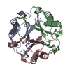
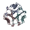
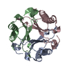
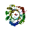
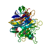
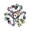
| Title | Kinetic and structural characterization of the 4-oxalocrotonate tautomerase isozymes from Methylibium petroleiphilum | ||||||
 Components Components | 4-oxalocrotonate isomerase protein | ||||||
 Keywords Keywords | ISOMERASE / alpha/beta fold / Tautomerase | ||||||
| Function / homology |  Function and homology information Function and homology informationIsomerases; Intramolecular oxidoreductases; Interconverting keto- and enol-groups / isomerase activity Similarity search - Function | ||||||
| Biological species |  Methylibium petroleiphilum (bacteria) Methylibium petroleiphilum (bacteria) | ||||||
| Method |  X-RAY DIFFRACTION / MOLECULAR REPLACEMENT / Resolution: 1.57 Å X-RAY DIFFRACTION / MOLECULAR REPLACEMENT / Resolution: 1.57 Å | ||||||
 Authors Authors | Terrell, C.R. / Hoffman, D.W. / Whitman, C.P. | ||||||
 Citation Citation | Journal: Arch.Biochem.Biophys. / Year: 2013 Title: Structural and kinetic characterization of two 4-oxalocrotonate tautomerases in Methylibium petroleiphilum strain PM1. Authors: Terrell, C.R. / Burks, E.A. / Whitman, C.P. / Hoffman, D.W. | ||||||
| History |
|
- Structure visualization
| Structure viewer | Molecule: MolmilJmol/JSmol |
|---|
- Downloads & links
-Download
| PDBx/mmCIF format | 4faz.cif.gz | 49 KB | Display | PDBx/mmCIF format |
|---|---|---|---|---|
| PDB format | pdb4faz.ent.gz | 35.5 KB | Display | PDB format |
| PDBx/mmJSON format | 4faz.json.gz | Tree view | PDBx/mmJSON format | |
| Others |  Other downloads Other downloads |
-Validation report
| Arichive directory | https://data.pdbj.org/pub/pdb/validation_reports/fa/4fazftp://data.pdbj.org/pub/pdb/validation_reports/fa/4faz | HTTPS FTP |
|---|
-Related structure data
| Related structure data | 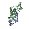 4fdxC 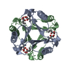 1bjpS C: citing same article ( S: Starting model for refinement |
|---|---|
| Similar structure data |

-Links
 PDBj
PDBj- Assembly
| Deposited unit | 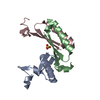
| ||||||||
|---|---|---|---|---|---|---|---|---|---|
| 1 | 
| ||||||||
| Unit cell |
|
-Components
| #1: Protein | Mass: 6913.975 Da / Num. of mol.: 3 Source method: isolated from a genetically manipulated source Source: (gene. exp.) Methylibium petroleiphilum (bacteria) / Strain: PM1 / Gene: Mpe_A2265 / Plasmid: pET24 / Production host: References: UniProt: A2SI32, Isomerases; Intramolecular oxidoreductases; Interconverting keto- and enol-groups #2: Chemical | ChemComp-SO4 / |   Mass: 96.063 Da / Num. of mol.: 1 / Source method: obtained synthetically / Formula: SO4 Mass: 96.063 Da / Num. of mol.: 1 / Source method: obtained synthetically / Formula: SO4#3: Water | ChemComp-HOH / |  Mass: 18.015 Da / Num. of mol.: 61 / Source method: isolated from a natural source / Formula: H2O Mass: 18.015 Da / Num. of mol.: 61 / Source method: isolated from a natural source / Formula: H2O |
|---|
-Experimental details
-Experiment
| Experiment | Method: X-RAY DIFFRACTION / Number of used crystals: 1 |
|---|
- Sample preparation
| Crystal | Density Matthews: 1.93 Å3/Da / Density % sol: 36.27 % |
|---|---|
| Crystal grow | Temperature: 298 K / Method: vapor diffusion, hanging drop / pH: 6.5 Details: 200 mM tri-Ammonium Citrate, 10% PEG 3350, pH 6.5, VAPOR DIFFUSION, HANGING DROP, temperature 298K |
-Data collection
| Diffraction | Mean temperature: 100 K |
|---|---|
| Diffraction source | Source: ROTATING ANODE / Type: RIGAKU MICROMAX-007 / Wavelength: 1.5418 Å |
| Detector | Type: RIGAKU RAXIS IV++ / Detector: IMAGE PLATE / Date: Aug 21, 2011 |
| Radiation | Protocol: SINGLE WAVELENGTH / Monochromatic (M) / Laue (L): M / Scattering type: x-ray |
| Radiation wavelength | Wavelength: 1.5418 Å / Relative weight: 1 |
| Reflection | Resolution: 1.57→47.22 Å / Num. all: 24295 / Num. obs: 20889 / % possible obs: 98.5 % / Observed criterion σ(F): 0 / Observed criterion σ(I): 0 |
| Reflection shell | Resolution: 1.57→1.61 Å / % possible all: 98.5 |
- Processing
| Software |
| |||||||||||||||||||||||||||||||||||||||||||||
|---|---|---|---|---|---|---|---|---|---|---|---|---|---|---|---|---|---|---|---|---|---|---|---|---|---|---|---|---|---|---|---|---|---|---|---|---|---|---|---|---|---|---|---|---|---|---|
| Refinement | Method to determine structure: MOLECULAR REPLACEMENT Starting model: PDB entry 1BJP Resolution: 1.57→47.22 Å / Cor.coef. Fo:Fc: 0.951 / Cor.coef. Fo:Fc free: 0.934 / SU B: 1.955 / SU ML: 0.072 / Cross valid method: THROUGHOUT / ESU R: 0.119 / ESU R Free: 0.12 / Stereochemistry target values: MAXIMUM LIKELIHOOD / Details: HYDROGENS HAVE BEEN USED IF PRESENT IN THE INPUT
| |||||||||||||||||||||||||||||||||||||||||||||
| Solvent computation | Ion probe radii: 0.8 Å / Shrinkage radii: 0.8 Å / VDW probe radii: 1.2 Å / Solvent model: MASK | |||||||||||||||||||||||||||||||||||||||||||||
| Displacement parameters | Biso mean: 26.871 Å2
| |||||||||||||||||||||||||||||||||||||||||||||
| Refinement step | Cycle: LAST / Resolution: 1.57→47.22 Å
| |||||||||||||||||||||||||||||||||||||||||||||
| Refine LS restraints |
| |||||||||||||||||||||||||||||||||||||||||||||
| LS refinement shell | Resolution: 1.57→1.61 Å / Total num. of bins used: 20
|
