 Movie
Movie Controller


+ Open data

- Basic information
| Entry | Database: PDB / ID: 4fak | ||||||
|---|---|---|---|---|---|---|---|
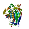
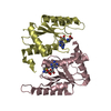
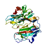
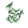
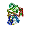
| Title | Crystal Structure of OrfX in Complex with S-Adenosylmethionine | ||||||
 Components Components | Ribosomal RNA large subunit methyltransferase H | ||||||
 Keywords Keywords | TRANSFERASE / RIBOSOMAL PROTEIN / alpha/beta methyltransferase Rossmann fold / rRNA methylation / AdoMet / rRNA | ||||||
| Function / homology |  Function and homology information Function and homology information23S rRNA (pseudouridine1915-N3)-methyltransferase / rRNA (pseudouridine-N3-)-methyltransferase activity / cytoplasm Similarity search - Function | ||||||
| Biological species |   Staphylococcus aureus (bacteria) Staphylococcus aureus (bacteria) | ||||||
| Method |  X-RAY DIFFRACTION / MOLECULAR REPLACEMENT / Resolution: 1.7 Å X-RAY DIFFRACTION / MOLECULAR REPLACEMENT / Resolution: 1.7 Å | ||||||
 Authors Authors | Safo, M.K. / Musayev, F.N. / Boundy, S. / Archer, G.L. / Rife, J.P. / O'Farrell, H.C. | ||||||
 Citation Citation | Journal: J.Biol.Chem. / Year: 2013 Title: Characterization of the Staphylococcus aureus rRNA Methyltransferase Encoded by orfX, the Gene Containing the Staphylococcal Chromosome Cassette mec (SCCmec) Insertion Site. Authors: Boundy, S. / Safo, M.K. / Wang, L. / Musayev, F.N. / O'Farrell, H.C. / Rife, J.P. / Archer, G.L. | ||||||
| History |
|
- Structure visualization
| Structure viewer | Molecule: MolmilJmol/JSmol |
|---|
- Downloads & links
-Download
| PDBx/mmCIF format | 4fak.cif.gz | 53.4 KB | Display | PDBx/mmCIF format |
|---|---|---|---|---|
| PDB format | pdb4fak.ent.gz | 36.2 KB | Display | PDB format |
| PDBx/mmJSON format | 4fak.json.gz | Tree view | PDBx/mmJSON format | |
| Others |  Other downloads Other downloads |
-Validation report
| Arichive directory | https://data.pdbj.org/pub/pdb/validation_reports/fa/4fakftp://data.pdbj.org/pub/pdb/validation_reports/fa/4fak | HTTPS FTP |
|---|
-Related structure data
| Related structure data | 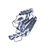 1vh0S S: Starting model for refinement |
|---|---|
| Similar structure data |


-Links
 PDBj
PDBj
- Assembly
| Deposited unit | 
| |||||||||||||||
|---|---|---|---|---|---|---|---|---|---|---|---|---|---|---|---|---|
| 1 | 
| |||||||||||||||
| Unit cell |
| |||||||||||||||
| Components on special symmetry positions |
|
-Components
| #1: Protein | Mass: 18752.818 Da / Num. of mol.: 1 Source method: isolated from a genetically manipulated source Source: (gene. exp.) Staphylococcus aureus (bacteria) / Strain: RN450M / Gene: orfx, rlmH / Plasmid: pET30a / Production host: References: UniProt: P0C1V0, 23S rRNA (pseudouridine1915-N3)-methyltransferase |
|---|---|
| #2: Chemical | ChemComp-SAM /   Mass: 398.437 Da / Num. of mol.: 1 / Source method: obtained synthetically / Formula: C15H22N6O5S Mass: 398.437 Da / Num. of mol.: 1 / Source method: obtained synthetically / Formula: C15H22N6O5S |
| #3: Chemical | ChemComp-PG4 /   Mass: 194.226 Da / Num. of mol.: 1 / Source method: obtained synthetically / Formula: C8H18O5 / Comment: precipitant*YM Mass: 194.226 Da / Num. of mol.: 1 / Source method: obtained synthetically / Formula: C8H18O5 / Comment: precipitant*YM |
| #4: Chemical | ChemComp-PO4 /   Mass: 94.971 Da / Num. of mol.: 1 / Source method: obtained synthetically / Formula: PO4 Mass: 94.971 Da / Num. of mol.: 1 / Source method: obtained synthetically / Formula: PO4 |
| #5: Water | ChemComp-HOH /  Mass: 18.015 Da / Num. of mol.: 177 / Source method: isolated from a natural source / Formula: H2O Mass: 18.015 Da / Num. of mol.: 177 / Source method: isolated from a natural source / Formula: H2O |
-Experimental details
-Experiment
| Experiment | Method: X-RAY DIFFRACTION / Number of used crystals: 1 |
|---|
- Sample preparation
| Crystal | Density Matthews: 2.44 Å3/Da / Density % sol: 49.51 % |
|---|---|
| Crystal grow | Temperature: 290 K / Method: vapor diffusion, hanging drop / pH: 6 Details: 20% PEG 550 MME, 5 mM ZnSO4, 0.1M Na 2-(N-morpholino) ethanesulfonate, pH 6.0, VAPOR DIFFUSION, HANGING DROP, temperature 290K |
-Data collection
| Diffraction | Mean temperature: 100 K |
|---|---|
| Diffraction source | Source: ROTATING ANODE / Type: RIGAKU MICROMAX-007 / Wavelength: 1.5418 Å |
| Detector | Type: RIGAKU RAXIS IV++ / Detector: IMAGE PLATE / Date: Feb 1, 2010 / Details: mirrors |
| Radiation | Monochromator: Mirrors / Protocol: SINGLE WAVELENGTH / Monochromatic (M) / Laue (L): M / Scattering type: x-ray |
| Radiation wavelength | Wavelength: 1.5418 Å / Relative weight: 1 |
| Reflection | Resolution: 1.7→28.6 Å / Num. all: 19664 / Num. obs: 20528 / % possible obs: 95.8 % / Observed criterion σ(F): 0 / Observed criterion σ(I): 0 / Redundancy: 8.4 % / Biso Wilson estimate: 26 Å2 / Rmerge(I) obs: 0.059 / Net I/σ(I): 19.5 |
| Reflection shell | Resolution: 1.7→1.76 Å / Rmerge(I) obs: 0.347 / Mean I/σ(I) obs: 5.4 / Num. unique all: 1862 / % possible all: 93.8 |
- Processing
| Software |
| ||||||||||||||||||||||||||||||||||||||||||||||||||||||||||||||||||||||||||||||||
|---|---|---|---|---|---|---|---|---|---|---|---|---|---|---|---|---|---|---|---|---|---|---|---|---|---|---|---|---|---|---|---|---|---|---|---|---|---|---|---|---|---|---|---|---|---|---|---|---|---|---|---|---|---|---|---|---|---|---|---|---|---|---|---|---|---|---|---|---|---|---|---|---|---|---|---|---|---|---|---|---|---|
| Refinement | Method to determine structure: MOLECULAR REPLACEMENT Starting model: PDB ENTRY 1VH0 Resolution: 1.7→26.43 Å / Rfactor Rfree error: 0.008 / Data cutoff high absF: 1372577.11 / Data cutoff low absF: 0 / Isotropic thermal model: RESTRAINED / Cross valid method: THROUGHOUT / σ(F): 0 / Stereochemistry target values: Engh & Huber
| ||||||||||||||||||||||||||||||||||||||||||||||||||||||||||||||||||||||||||||||||
| Solvent computation | Solvent model: FLAT MODEL / Bsol: 54.9051 Å2 / ksol: 0.355692 e/Å3 | ||||||||||||||||||||||||||||||||||||||||||||||||||||||||||||||||||||||||||||||||
| Displacement parameters | Biso mean: 30.8 Å2
| ||||||||||||||||||||||||||||||||||||||||||||||||||||||||||||||||||||||||||||||||
| Refine analyze |
| ||||||||||||||||||||||||||||||||||||||||||||||||||||||||||||||||||||||||||||||||
| Refinement step | Cycle: LAST / Resolution: 1.7→26.43 Å
| ||||||||||||||||||||||||||||||||||||||||||||||||||||||||||||||||||||||||||||||||
| Refine LS restraints |
| ||||||||||||||||||||||||||||||||||||||||||||||||||||||||||||||||||||||||||||||||
| Refine LS restraints NCS | NCS model details: NONE | ||||||||||||||||||||||||||||||||||||||||||||||||||||||||||||||||||||||||||||||||
| LS refinement shell | Resolution: 1.7→1.76 Å / Rfactor Rfree error: 0.038 / Total num. of bins used: 10
|
