 Movie
Movie Controller

[English] 日本語
 Yorodumi
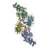
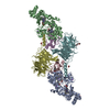
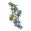
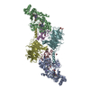
- PDB-4fa1: Crystal Structure of WT MauG in Complex with Pre-Methylamine Dehy... -
+ Open data

- Basic information
| Entry | Database: PDB / ID: 4fa1 | ||||||
|---|---|---|---|---|---|---|---|
| Title | Crystal Structure of WT MauG in Complex with Pre-Methylamine Dehydrogenase Aged 130 Days. | ||||||
 Components Components |
| ||||||
 Keywords Keywords | Oxidoreductase/Electron transport / tryptophan tryptophylquinone / Oxidoreductase-Electron Transfer complex / Oxidoreductase-Electron transport complex | ||||||
| Function / homology |  Function and homology information Function and homology informationmethylamine dehydrogenase (amicyanin) / methylamine dehydrogenase (amicyanin) activity / methylamine metabolic process / aliphatic amine dehydrogenase activity / amine metabolic process / cytochrome-c peroxidase activity / Oxidoreductases / outer membrane-bounded periplasmic space / electron transfer activity / periplasmic space ...methylamine dehydrogenase (amicyanin) / methylamine dehydrogenase (amicyanin) activity / methylamine metabolic process / aliphatic amine dehydrogenase activity / amine metabolic process / cytochrome-c peroxidase activity / Oxidoreductases / outer membrane-bounded periplasmic space / electron transfer activity / periplasmic space / heme binding / metal ion binding Similarity search - Function | ||||||
| Biological species |  Paracoccus denitrificans (bacteria) Paracoccus denitrificans (bacteria) | ||||||
| Method |  X-RAY DIFFRACTION / SYNCHROTRON / FOURIER SYNTHESIS / Resolution: 2.18 Å X-RAY DIFFRACTION / SYNCHROTRON / FOURIER SYNTHESIS / Resolution: 2.18 Å | ||||||
 Authors Authors | Yukl, E.T. / Wilmot, C.M. | ||||||
 Citation Citation | Journal: Proc.Natl.Acad.Sci.USA / Year: 2013 Title: Diradical intermediate within the context of tryptophan tryptophylquinone biosynthesis. Authors: Yukl, E.T. / Liu, F. / Krzystek, J. / Shin, S. / Jensen, L.M. / Davidson, V.L. / Wilmot, C.M. / Liu, A. | ||||||
| History |
|
- Structure visualization
| Structure viewer | Molecule: MolmilJmol/JSmol |
|---|
- Downloads & links
-Download
| PDBx/mmCIF format | 4fa1.cif.gz | 699.6 KB | Display | PDBx/mmCIF format |
|---|---|---|---|---|
| PDB format | pdb4fa1.ent.gz | 577.1 KB | Display | PDB format |
| PDBx/mmJSON format | 4fa1.json.gz | Tree view | PDBx/mmJSON format | |
| Others |  Other downloads Other downloads |
-Validation report
| Arichive directory | https://data.pdbj.org/pub/pdb/validation_reports/fa/4fa1ftp://data.pdbj.org/pub/pdb/validation_reports/fa/4fa1 | HTTPS FTP |
|---|
-Related structure data
| Related structure data |  4fa4C 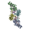 4fa5C  4fa9C 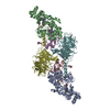 4fanC 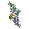 4favC 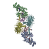 4fb1C 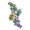 3l4mS S: Starting model for refinement C: citing same article ( |
|---|---|
| Similar structure data |
-Links
 PDBj
PDBj


- Assembly
| Deposited unit | 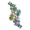
| ||||||||
|---|---|---|---|---|---|---|---|---|---|
| 1 |
| ||||||||
| Unit cell |
| ||||||||
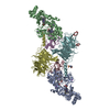
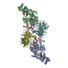
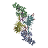
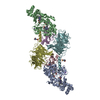
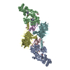
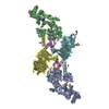
| Details | Pre-methylamine dehydrogenase is a heterotetramer of 2 light and 2 heavy chains. Two copies of MauG are bound to each pre-MADH tetramer, making for a 6-chain complex in the asymmetric unit. |
-Components
-Protein / Antibody / Methylamine dehydrogenase ... , 3 types, 6 molecules ABCEDF
| #1: Protein | Mass: 41146.629 Da / Num. of mol.: 2 Source method: isolated from a genetically manipulated source Source: (gene. exp.) Paracoccus denitrificans (bacteria) / Strain: PD1222 / Gene: mauG, Pden_4736 / Production host: Paracoccus denitrificans (bacteria) / References: UniProt: Q51658, Oxidoreductases#2: Antibody | Mass: 15039.577 Da / Num. of mol.: 2 Source method: isolated from a genetically manipulated source Source: (gene. exp.) Paracoccus denitrificans (bacteria) / Strain: PD1222 / Gene: mauA / Production host: Rhodobacter sphaeroides (bacteria)References: UniProt: P22619, methylamine dehydrogenase (amicyanin) #3: Protein | Mass: 42321.152 Da / Num. of mol.: 2 Source method: isolated from a genetically manipulated source Source: (gene. exp.) Paracoccus denitrificans (bacteria) / Strain: PD1222 / Gene: Pden_4730 / Production host: Rhodobacter sphaeroides (bacteria) / References: UniProt: A1BB97, EC: 1.4.99.3 |
|---|
-Non-polymers , 7 types, 1023 molecules 


| #4: Chemical |  Mass: 40.078 Da / Num. of mol.: 2 / Source method: obtained synthetically / Formula: Ca Mass: 40.078 Da / Num. of mol.: 2 / Source method: obtained synthetically / Formula: Ca#5: Chemical | ChemComp-HEC /  Mass: 618.503 Da / Num. of mol.: 4 / Source method: obtained synthetically / Formula: C34H34FeN4O4 Mass: 618.503 Da / Num. of mol.: 4 / Source method: obtained synthetically / Formula: C34H34FeN4O4#6: Chemical |  Mass: 22.990 Da / Num. of mol.: 2 / Source method: obtained synthetically / Formula: Na Mass: 22.990 Da / Num. of mol.: 2 / Source method: obtained synthetically / Formula: Na#7: Chemical |  Mass: 62.068 Da / Num. of mol.: 2 / Source method: obtained synthetically / Formula: C2H6O2 Mass: 62.068 Da / Num. of mol.: 2 / Source method: obtained synthetically / Formula: C2H6O2#8: Chemical | ChemComp-ACT / |  Mass: 59.044 Da / Num. of mol.: 1 / Source method: obtained synthetically / Formula: C2H3O2 Mass: 59.044 Da / Num. of mol.: 1 / Source method: obtained synthetically / Formula: C2H3O2#9: Chemical | ChemComp-MES / |  Mass: 195.237 Da / Num. of mol.: 1 / Source method: obtained synthetically / Formula: C6H13NO4S / Comment: pH buffer*YM Mass: 195.237 Da / Num. of mol.: 1 / Source method: obtained synthetically / Formula: C6H13NO4S / Comment: pH buffer*YM#10: Water | ChemComp-HOH / | Mass: 18.015 Da / Num. of mol.: 1011 / Source method: isolated from a natural source / Formula: H2O |
|---|
-Experimental details
-Experiment
| Experiment | Method: X-RAY DIFFRACTION / Number of used crystals: 1 |
|---|
- Sample preparation
| Crystal | Density Matthews: 2.28 Å3/Da / Density % sol: 45.94 % |
|---|---|
| Crystal grow | Temperature: 293 K / Method: vapor diffusion, hanging drop / pH: 6.4 Details: 0.1M MES pH 6.4, 0.1M sodium acetate, 22-26 % w/v PEG 8000, VAPOR DIFFUSION, HANGING DROP, temperature 293K |
-Data collection
| Diffraction | Mean temperature: 100 K |
|---|---|
| Diffraction source | Source: SYNCHROTRON / Site: APS  / Beamline: 23-ID-B / Wavelength: 1.0332 Å / Beamline: 23-ID-B / Wavelength: 1.0332 Å |
| Detector | Type: MARMOSAIC 300 mm CCD / Detector: CCD / Date: Feb 18, 2011 |
| Radiation | Monochromator: SI(111) DOUBLE CRYSTAL MONOCHROMATOR / Protocol: SINGLE WAVELENGTH / Monochromatic (M) / Laue (L): M / Scattering type: x-ray |
| Radiation wavelength | Wavelength: 1.0332 Å / Relative weight: 1 |
| Reflection | Resolution: 2.18→50 Å / Num. all: 92667 / Num. obs: 85956 / % possible obs: 92.8 % / Observed criterion σ(F): 0 / Observed criterion σ(I): -3 / Redundancy: 2.1 % / Rmerge(I) obs: 0.071 / Net I/σ(I): 11.58 |
| Reflection shell | Resolution: 2.18→2.22 Å / Redundancy: 2 % / Rmerge(I) obs: 0.353 / Mean I/σ(I) obs: 2.03 / % possible all: 69.3 |
- Processing
| Software |
| ||||||||||||||||||||||||||||||||||||||||||||||||||||||||||||||||||||||||||||||||||||||||||||||||||||||||||||||||||||||||||||||||||||||||||||||||||||||||||||||||||||||||||
|---|---|---|---|---|---|---|---|---|---|---|---|---|---|---|---|---|---|---|---|---|---|---|---|---|---|---|---|---|---|---|---|---|---|---|---|---|---|---|---|---|---|---|---|---|---|---|---|---|---|---|---|---|---|---|---|---|---|---|---|---|---|---|---|---|---|---|---|---|---|---|---|---|---|---|---|---|---|---|---|---|---|---|---|---|---|---|---|---|---|---|---|---|---|---|---|---|---|---|---|---|---|---|---|---|---|---|---|---|---|---|---|---|---|---|---|---|---|---|---|---|---|---|---|---|---|---|---|---|---|---|---|---|---|---|---|---|---|---|---|---|---|---|---|---|---|---|---|---|---|---|---|---|---|---|---|---|---|---|---|---|---|---|---|---|---|---|---|---|---|---|---|
| Refinement | Method to determine structure: FOURIER SYNTHESIS Starting model: 3L4M Resolution: 2.18→37.17 Å / Cor.coef. Fo:Fc: 0.967 / Cor.coef. Fo:Fc free: 0.93 / SU B: 14.216 / SU ML: 0.161 / Cross valid method: THROUGHOUT / ESU R: 0.29 / ESU R Free: 0.219 / Stereochemistry target values: MAXIMUM LIKELIHOOD / Details: HYDROGENS HAVE BEEN ADDED IN THE RIDING POSITIONS
| ||||||||||||||||||||||||||||||||||||||||||||||||||||||||||||||||||||||||||||||||||||||||||||||||||||||||||||||||||||||||||||||||||||||||||||||||||||||||||||||||||||||||||
| Solvent computation | Ion probe radii: 0.8 Å / Shrinkage radii: 0.8 Å / VDW probe radii: 1.4 Å / Solvent model: BABINET MODEL WITH MASK | ||||||||||||||||||||||||||||||||||||||||||||||||||||||||||||||||||||||||||||||||||||||||||||||||||||||||||||||||||||||||||||||||||||||||||||||||||||||||||||||||||||||||||
| Displacement parameters | Biso mean: 37.752 Å2
| ||||||||||||||||||||||||||||||||||||||||||||||||||||||||||||||||||||||||||||||||||||||||||||||||||||||||||||||||||||||||||||||||||||||||||||||||||||||||||||||||||||||||||
| Refinement step | Cycle: LAST / Resolution: 2.18→37.17 Å
| ||||||||||||||||||||||||||||||||||||||||||||||||||||||||||||||||||||||||||||||||||||||||||||||||||||||||||||||||||||||||||||||||||||||||||||||||||||||||||||||||||||||||||
| Refine LS restraints |
| ||||||||||||||||||||||||||||||||||||||||||||||||||||||||||||||||||||||||||||||||||||||||||||||||||||||||||||||||||||||||||||||||||||||||||||||||||||||||||||||||||||||||||
| LS refinement shell | Resolution: 2.18→2.236 Å / Total num. of bins used: 20
| ||||||||||||||||||||||||||||||||||||||||||||||||||||||||||||||||||||||||||||||||||||||||||||||||||||||||||||||||||||||||||||||||||||||||||||||||||||||||||||||||||||||||||
| Refinement TLS params. | Method: refined / Refine-ID: X-RAY DIFFRACTION
| ||||||||||||||||||||||||||||||||||||||||||||||||||||||||||||||||||||||||||||||||||||||||||||||||||||||||||||||||||||||||||||||||||||||||||||||||||||||||||||||||||||||||||
| Refinement TLS group |
|
