 Movie
Movie Controller

[English] 日本語
 Yorodumi
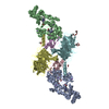
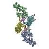
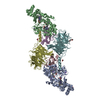
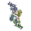
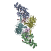
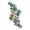
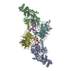
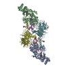
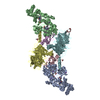
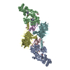
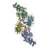
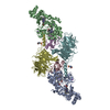
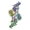
- PDB-3l4m: Crystal Structure of the MauG/pre-Methylamine Dehydrogenase Complex. -
+ Open data

- Basic information
| Entry | Database: PDB / ID: 3l4m | ||||||
|---|---|---|---|---|---|---|---|
| Title | Crystal Structure of the MauG/pre-Methylamine Dehydrogenase Complex. | ||||||
 Components Components |
| ||||||
 Keywords Keywords | OXIDOREDUCTASE/ELECTRON TRANSPORT / MauG / methylamine dehydrogenase / quinone cofactor / TTQ / His-Tyr heme / Electron transport / c-heme / Iron / Metal-binding / Oxidoreductase / Transport / Disulfide bond / OXIDOREDUCTASE-ELECTRON TRANSPORT complex | ||||||
| Function / homology |  Function and homology information Function and homology informationmethylamine dehydrogenase (amicyanin) / methylamine dehydrogenase (amicyanin) activity / methylamine metabolic process / aliphatic amine dehydrogenase activity / amine metabolic process / cytochrome-c peroxidase activity / Oxidoreductases / outer membrane-bounded periplasmic space / electron transfer activity / periplasmic space ...methylamine dehydrogenase (amicyanin) / methylamine dehydrogenase (amicyanin) activity / methylamine metabolic process / aliphatic amine dehydrogenase activity / amine metabolic process / cytochrome-c peroxidase activity / Oxidoreductases / outer membrane-bounded periplasmic space / electron transfer activity / periplasmic space / heme binding / metal ion binding Similarity search - Function | ||||||
| Biological species |  Paracoccus denitrificans (bacteria) Paracoccus denitrificans (bacteria) | ||||||
| Method |  X-RAY DIFFRACTION / SYNCHROTRON / MOLECULAR REPLACEMENT / molecular replacement / Resolution: 2.02 Å X-RAY DIFFRACTION / SYNCHROTRON / MOLECULAR REPLACEMENT / molecular replacement / Resolution: 2.02 Å | ||||||
 Authors Authors | Jensen, L.M.R. / Wilmot, C.M. | ||||||
 Citation Citation | Journal: Science / Year: 2010 Title: In crystallo posttranslational modification within a MauG/pre-methylamine dehydrogenase complex. Authors: Jensen, L.M. / Sanishvili, R. / Davidson, V.L. / Wilmot, C.M. | ||||||
| History |
|
- Structure visualization
| Structure viewer | Molecule: MolmilJmol/JSmol |
|---|
- Downloads & links
-Download
| PDBx/mmCIF format | 3l4m.cif.gz | 377.5 KB | Display | PDBx/mmCIF format |
|---|---|---|---|---|
| PDB format | pdb3l4m.ent.gz | 300.4 KB | Display | PDB format |
| PDBx/mmJSON format | 3l4m.json.gz | Tree view | PDBx/mmJSON format | |
| Others |  Other downloads Other downloads |
-Validation report
| Arichive directory | https://data.pdbj.org/pub/pdb/validation_reports/l4/3l4mftp://data.pdbj.org/pub/pdb/validation_reports/l4/3l4m | HTTPS FTP |
|---|
-Related structure data
| Related structure data |  3l4oC 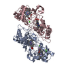 1iqcS  1mdaS C: citing same article ( S: Starting model for refinement |
|---|---|
| Similar structure data |


-Links
 PDBj
PDBj


- Assembly
| Deposited unit | 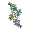
| ||||||||
|---|---|---|---|---|---|---|---|---|---|
| 1 |
| ||||||||
| Unit cell |
|
-Components
-Protein / Antibody / Methylamine dehydrogenase ... , 3 types, 6 molecules ABCEDF
| #1: Protein | Mass: 41146.629 Da / Num. of mol.: 2 Source method: isolated from a genetically manipulated source Source: (gene. exp.) Paracoccus denitrificans (bacteria) / Strain: Pd 1222 / Gene: mauG / Production host: Paracoccus denitrificans (bacteria) / References: UniProt: Q51658, Oxidoreductases#2: Antibody | Mass: 15025.595 Da / Num. of mol.: 2 Fragment: Beta chain of immature methylamine dehydrogenase (preMADH) Mutation: Trp57 is hydroxylated at C7 Source method: isolated from a genetically manipulated source Details: Immature MADH (preMADH) was produced in the absence of the mauG gene. Trp57 is monohydroxylated at the C7 position. Source: (gene. exp.) Paracoccus denitrificans (bacteria) / Strain: Pd 1222 / Gene: mauA / Production host: Rhodobacter sphaeroides (bacteria)References: UniProt: P22619, UniProt: A1BBA0*PLUS, EC: 1.4.99.3 #3: Protein | Mass: 42449.277 Da / Num. of mol.: 2 Source method: isolated from a genetically manipulated source Details: Immature MADH (preMADH) was produced in the absence of the mauG gene. The alpha subunit has the wild-type sequence. Source: (gene. exp.) Paracoccus denitrificans (bacteria) / Strain: Pd 1222 / Gene: Pden_4730 / Production host: Rhodobacter sphaeroides (bacteria) / References: UniProt: A1BB97, EC: 1.4.99.3 |
|---|
-Non-polymers , 6 types, 1313 molecules 


| #4: Chemical |  Mass: 40.078 Da / Num. of mol.: 2 / Source method: obtained synthetically / Formula: Ca Mass: 40.078 Da / Num. of mol.: 2 / Source method: obtained synthetically / Formula: Ca#5: Chemical | ChemComp-HEC /  Mass: 618.503 Da / Num. of mol.: 4 / Source method: obtained synthetically / Formula: C34H34FeN4O4 Mass: 618.503 Da / Num. of mol.: 4 / Source method: obtained synthetically / Formula: C34H34FeN4O4#6: Chemical | ChemComp-1PE / |  Mass: 238.278 Da / Num. of mol.: 1 / Source method: obtained synthetically / Formula: C10H22O6 / Comment: precipitant*YM Mass: 238.278 Da / Num. of mol.: 1 / Source method: obtained synthetically / Formula: C10H22O6 / Comment: precipitant*YM#7: Chemical | ChemComp-PG4 / |  Mass: 194.226 Da / Num. of mol.: 1 / Source method: obtained synthetically / Formula: C8H18O5 / Comment: precipitant*YM Mass: 194.226 Da / Num. of mol.: 1 / Source method: obtained synthetically / Formula: C8H18O5 / Comment: precipitant*YM#8: Chemical | ChemComp-ACT / |  Mass: 59.044 Da / Num. of mol.: 1 / Source method: obtained synthetically / Formula: C2H3O2 Mass: 59.044 Da / Num. of mol.: 1 / Source method: obtained synthetically / Formula: C2H3O2#9: Water | ChemComp-HOH / | Mass: 18.015 Da / Num. of mol.: 1304 / Source method: isolated from a natural source / Formula: H2O |
|---|
-Details
| Has protein modification | Y |
|---|
-Experimental details
-Experiment
| Experiment | Method: X-RAY DIFFRACTION / Number of used crystals: 1 |
|---|
- Sample preparation
| Crystal | Density Matthews: 2.29 Å3/Da / Density % sol: 46.33 % |
|---|---|
| Crystal grow | Temperature: 293 K / Method: vapor diffusion, hanging drop / pH: 6.4 Details: 0.1M MES pH 6.4, 0.1M sodium acetate, 24-30 % w/v PEG 8000, vapor diffusion, hanging drop, temperature 293K |
-Data collection
| Diffraction | Mean temperature: 100 K |
|---|---|
| Diffraction source | Source: SYNCHROTRON / Site: APS  / Beamline: 23-ID-B / Wavelength: 1.02665 Å / Beamline: 23-ID-B / Wavelength: 1.02665 Å |
| Detector | Type: MARMOSAIC 300 mm CCD / Detector: CCD / Date: Nov 25, 2008 / Details: BIOMORPH MIRRORS (KIRKPATRICK-BAEZ CONFIGURATION) |
| Radiation | Monochromator: Si(111) Double crystal monochromator / Protocol: SINGLE WAVELENGTH / Monochromatic (M) / Laue (L): M / Scattering type: x-ray |
| Radiation wavelength | Wavelength: 1.02665 Å / Relative weight: 1 |
| Reflection | Resolution: 2.01→50 Å / Num. all: 113264 / Num. obs: 105427 / % possible obs: 93.1 % / Observed criterion σ(F): 0 / Observed criterion σ(I): -3 / Redundancy: 2.3 % / Biso Wilson estimate: 27.03 Å2 / Rmerge(I) obs: 0.067 / Rsym value: 0.067 / Net I/σ(I): 10.74 |
| Reflection shell | Resolution: 2.01→2.08 Å / Redundancy: 1.7 % / Rmerge(I) obs: 0.176 / Mean I/σ(I) obs: 4.3 / Num. unique all: 11318 / % possible all: 68.6 |
-Phasing
| Phasing | Method: molecular replacement |
|---|
- Processing
| Software |
| |||||||||||||||||||||||||||||||||||||||||||||||||||||||||||||||||||||||||||||||||||||||||||||||||||||||||||||||||||||||||||||||||||||||||||||||||||||||||||||||||||||||||||||||
|---|---|---|---|---|---|---|---|---|---|---|---|---|---|---|---|---|---|---|---|---|---|---|---|---|---|---|---|---|---|---|---|---|---|---|---|---|---|---|---|---|---|---|---|---|---|---|---|---|---|---|---|---|---|---|---|---|---|---|---|---|---|---|---|---|---|---|---|---|---|---|---|---|---|---|---|---|---|---|---|---|---|---|---|---|---|---|---|---|---|---|---|---|---|---|---|---|---|---|---|---|---|---|---|---|---|---|---|---|---|---|---|---|---|---|---|---|---|---|---|---|---|---|---|---|---|---|---|---|---|---|---|---|---|---|---|---|---|---|---|---|---|---|---|---|---|---|---|---|---|---|---|---|---|---|---|---|---|---|---|---|---|---|---|---|---|---|---|---|---|---|---|---|---|---|---|---|
| Refinement | Method to determine structure: MOLECULAR REPLACEMENT Starting model: 1mda, 1iqc Resolution: 2.02→44.49 Å / Cor.coef. Fo:Fc: 0.969 / Cor.coef. Fo:Fc free: 0.942 / WRfactor Rfree: 0.208 / WRfactor Rwork: 0.149 / Occupancy max: 1 / Occupancy min: 0.3 / FOM work R set: 0.893 / SU B: 7.499 / SU ML: 0.094 / SU R Cruickshank DPI: 0.171 / SU Rfree: 0.153 / TLS residual ADP flag: LIKELY RESIDUAL / Cross valid method: THROUGHOUT / σ(F): 0 / ESU R: 0.171 / ESU R Free: 0.153 / Stereochemistry target values: MAXIMUM LIKELIHOOD Details: HYDROGENS HAVE BEEN ADDED IN THE RIDING POSTIONS. U VALUES: RESIDUAL ONLY. The MADH model (1MDA) did not provide sufficient phasing to determine the structures of the MauG copies. The ...Details: HYDROGENS HAVE BEEN ADDED IN THE RIDING POSTIONS. U VALUES: RESIDUAL ONLY. The MADH model (1MDA) did not provide sufficient phasing to determine the structures of the MauG copies. The Chainsaw software within the CCP4i suite was used to reduce the CCP (1IQC) input (following sequence alignment with MauG) such that only strictly conserved residues near heme sites or else with secondary structure (alpha-helix) were retained as the input model. The missing MauG residues were added in an iterative manner by placing residues at termini of the discontinuous model peptide chains (as appropriate according to the electron-density map) and refining with successively more residues located for MauG.
| |||||||||||||||||||||||||||||||||||||||||||||||||||||||||||||||||||||||||||||||||||||||||||||||||||||||||||||||||||||||||||||||||||||||||||||||||||||||||||||||||||||||||||||||
| Solvent computation | Ion probe radii: 0.8 Å / Shrinkage radii: 0.8 Å / VDW probe radii: 1.4 Å / Solvent model: BABINET MODEL WITH MASK | |||||||||||||||||||||||||||||||||||||||||||||||||||||||||||||||||||||||||||||||||||||||||||||||||||||||||||||||||||||||||||||||||||||||||||||||||||||||||||||||||||||||||||||||
| Displacement parameters | Biso max: 60.58 Å2 / Biso mean: 15.169 Å2 / Biso min: 2 Å2
| |||||||||||||||||||||||||||||||||||||||||||||||||||||||||||||||||||||||||||||||||||||||||||||||||||||||||||||||||||||||||||||||||||||||||||||||||||||||||||||||||||||||||||||||
| Refinement step | Cycle: LAST / Resolution: 2.02→44.49 Å
| |||||||||||||||||||||||||||||||||||||||||||||||||||||||||||||||||||||||||||||||||||||||||||||||||||||||||||||||||||||||||||||||||||||||||||||||||||||||||||||||||||||||||||||||
| Refine LS restraints |
| |||||||||||||||||||||||||||||||||||||||||||||||||||||||||||||||||||||||||||||||||||||||||||||||||||||||||||||||||||||||||||||||||||||||||||||||||||||||||||||||||||||||||||||||
| LS refinement shell | Resolution: 2.021→2.073 Å / Total num. of bins used: 20
| |||||||||||||||||||||||||||||||||||||||||||||||||||||||||||||||||||||||||||||||||||||||||||||||||||||||||||||||||||||||||||||||||||||||||||||||||||||||||||||||||||||||||||||||
| Refinement TLS params. | Method: refined / Refine-ID: X-RAY DIFFRACTION
| |||||||||||||||||||||||||||||||||||||||||||||||||||||||||||||||||||||||||||||||||||||||||||||||||||||||||||||||||||||||||||||||||||||||||||||||||||||||||||||||||||||||||||||||
| Refinement TLS group |
|
