 Movie
Movie Controller

[English] 日本語
 Yorodumi
- PDB-3sxt: Crystal Structure of the Quinol Form of Methylamine Dehydrogenase... -
+ Open data

- Basic information
| Entry | Database: PDB / ID: 3sxt | ||||||
|---|---|---|---|---|---|---|---|
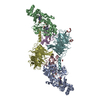
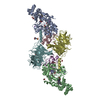
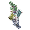
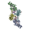
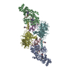
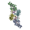
| Title | Crystal Structure of the Quinol Form of Methylamine Dehydrogenase in Complex with the Diferrous Form of MauG | ||||||
 Components Components |
| ||||||
 Keywords Keywords | OXIDOREDUCTASE/ELECTRON TRANSPORT / MauG / methylamine dehydrogenase / TTQ / c-heme / OXIDOREDUCTASE-ELECTRON TRANSPORT complex | ||||||
| Function / homology |  Function and homology information Function and homology informationmethylamine dehydrogenase (amicyanin) / methylamine dehydrogenase (amicyanin) activity / methylamine metabolic process / aliphatic amine dehydrogenase activity / amine metabolic process / cytochrome-c peroxidase activity / Oxidoreductases / outer membrane-bounded periplasmic space / electron transfer activity / periplasmic space ...methylamine dehydrogenase (amicyanin) / methylamine dehydrogenase (amicyanin) activity / methylamine metabolic process / aliphatic amine dehydrogenase activity / amine metabolic process / cytochrome-c peroxidase activity / Oxidoreductases / outer membrane-bounded periplasmic space / electron transfer activity / periplasmic space / heme binding / metal ion binding Similarity search - Function | ||||||
| Biological species |  Paracoccus denitrificans (bacteria) Paracoccus denitrificans (bacteria) | ||||||
| Method |  X-RAY DIFFRACTION / SYNCHROTRON / MOLECULAR REPLACEMENT / Resolution: 1.81 Å X-RAY DIFFRACTION / SYNCHROTRON / MOLECULAR REPLACEMENT / Resolution: 1.81 Å | ||||||
 Authors Authors | Jensen, L.M.R. / Wilmot, C.M. | ||||||
 Citation Citation | Journal: To be Published Title: Crystal Structure of the Quinol Form of Methylamine Dehydrogenase in Complex with the Diferrous Form of MauG Authors: Jensen, L.M.R. / Wilmot, C.M. | ||||||
| History |
|
- Structure visualization
| Structure viewer | Molecule: MolmilJmol/JSmol |
|---|
- Downloads & links
-Download
| PDBx/mmCIF format | 3sxt.cif.gz | 707.3 KB | Display | PDBx/mmCIF format |
|---|---|---|---|---|
| PDB format | pdb3sxt.ent.gz | 579.5 KB | Display | PDB format |
| PDBx/mmJSON format | 3sxt.json.gz | Tree view | PDBx/mmJSON format | |
| Others |  Other downloads Other downloads |
-Validation report
| Arichive directory | https://data.pdbj.org/pub/pdb/validation_reports/sx/3sxtftp://data.pdbj.org/pub/pdb/validation_reports/sx/3sxt | HTTPS FTP |
|---|
-Related structure data
| Related structure data | 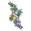 3l4mS S: Starting model for refinement |
|---|---|
| Similar structure data |

-Links
 PDBj
PDBj


- Assembly
| Deposited unit | 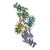
| ||||||||
|---|---|---|---|---|---|---|---|---|---|
| 1 |
| ||||||||
| Unit cell |
|
-Components
-Protein / Antibody / Methylamine dehydrogenase ... , 3 types, 6 molecules ABCEDF
| #1: Protein | Mass: 41146.629 Da / Num. of mol.: 2 / Fragment: UNP residues 21-387 Source method: isolated from a genetically manipulated source Source: (gene. exp.) Paracoccus denitrificans (bacteria) / Strain: Pd 1222 / Gene: mauG, Pden_4736 / Production host: Paracoccus denitrificans (bacteria) / References: UniProt: Q51658, Oxidoreductases#2: Antibody | Mass: 15041.593 Da / Num. of mol.: 2 / Fragment: UNP residues 58-188 Source method: isolated from a genetically manipulated source Details: Trp57 is quinol, with oxygen atoms at positions C6 and C7 of the indole ring Source: (gene. exp.) Paracoccus denitrificans (bacteria) / Strain: Pd 1222 / Gene: mauA / Production host: Rhodobacter sphaeroides (bacteria)References: UniProt: P22619, UniProt: A1BBA0*PLUS, EC: 1.4.99.3 #3: Protein | Mass: 42449.277 Da / Num. of mol.: 2 / Fragment: UNP residues 32-417 Source method: isolated from a genetically manipulated source Source: (gene. exp.) Paracoccus denitrificans (bacteria) / Strain: Pd 1222 / Gene: Pden_4730 / Production host: Rhodobacter sphaeroides (bacteria) / References: UniProt: A1BB97, EC: 1.4.99.3 |
|---|
-Non-polymers , 5 types, 1839 molecules 


| #4: Chemical |  Mass: 40.078 Da / Num. of mol.: 2 / Source method: obtained synthetically / Formula: Ca Mass: 40.078 Da / Num. of mol.: 2 / Source method: obtained synthetically / Formula: Ca#5: Chemical | ChemComp-NA /  Mass: 22.990 Da / Num. of mol.: 4 / Source method: obtained synthetically / Formula: Na Mass: 22.990 Da / Num. of mol.: 4 / Source method: obtained synthetically / Formula: Na#6: Chemical | ChemComp-HEC /  Mass: 618.503 Da / Num. of mol.: 4 / Source method: obtained synthetically / Formula: C34H34FeN4O4 Mass: 618.503 Da / Num. of mol.: 4 / Source method: obtained synthetically / Formula: C34H34FeN4O4#7: Chemical | ChemComp-EDO /  Mass: 62.068 Da / Num. of mol.: 5 / Source method: obtained synthetically / Formula: C2H6O2 Mass: 62.068 Da / Num. of mol.: 5 / Source method: obtained synthetically / Formula: C2H6O2#8: Water | ChemComp-HOH / | Mass: 18.015 Da / Num. of mol.: 1824 / Source method: isolated from a natural source / Formula: H2O |
|---|
-Details
| Has protein modification | Y |
|---|
-Experimental details
-Experiment
| Experiment | Method: X-RAY DIFFRACTION / Number of used crystals: 1 |
|---|
- Sample preparation
| Crystal | Density Matthews: 2.39 Å3/Da / Density % sol: 48.51 % |
|---|---|
| Crystal grow | Temperature: 293 K / pH: 6.4 Details: Drops contained 1uL protein with 3uL reservoir solution. WT-MauG and MADH were each reduced in an anaerobic glove box prior to preparing the protein mixture for crystallization. Protein ...Details: Drops contained 1uL protein with 3uL reservoir solution. WT-MauG and MADH were each reduced in an anaerobic glove box prior to preparing the protein mixture for crystallization. Protein mixture: 100uM reduced WT-MauG and 50uM reduced MADH in 10mM potassium phosphate pH7.5 with 2mM sodium dithionite. Reservoir solution contained: 22% w/v PEG 8000, 0.1M sodium acetate, 0.1M MES pH 6.4 and 2mM sodium dithionite. Crystallization was carried out in an anaerobic glove box at ambient temperature., VAPOR DIFFUSION, HANGING DROP, temperature 293K |
-Data collection
| Diffraction | Mean temperature: 100 K |
|---|---|
| Diffraction source | Source: SYNCHROTRON / Site: APS  / Beamline: 23-ID-B / Wavelength: 1.0332 / Beamline: 23-ID-B / Wavelength: 1.0332 |
| Detector | Type: MARMOSAIC 300 mm CCD / Detector: CCD / Date: Jun 29, 2011 / Details: BIOMORPH MIRRORS (KIRKPATRICK- BAEZ CONFIGURATION) |
| Radiation | Monochromator: SI(111) DOUBLE CRYSTAL MONOCHROMATOR / Protocol: SINGLE WAVELENGTH / Monochromatic (M) / Laue (L): M / Scattering type: x-ray |
| Radiation wavelength | Wavelength: 1.0332 Å / Relative weight: 1 |
| Reflection | Resolution: 1.81→50 Å / Num. obs: 164390 / % possible obs: 98.3 % / Observed criterion σ(I): -3 / Redundancy: 6.5 % / Rsym value: 0.093 / Net I/σ(I): 16.7 |
| Reflection shell | Resolution: 1.81→1.84 Å / Redundancy: 2.9 % / Mean I/σ(I) obs: 2.3 / Rsym value: 0.41 / % possible all: 80.9 |
- Processing
| Software |
| |||||||||||||||||||||||||||||||||||||||||||||||||||||||||||||||||||||||||||||||||||||||||||||||||||||||||||||||||||||||||||||||||||||||||||||||||||||||||||||||||||||||||||||||
|---|---|---|---|---|---|---|---|---|---|---|---|---|---|---|---|---|---|---|---|---|---|---|---|---|---|---|---|---|---|---|---|---|---|---|---|---|---|---|---|---|---|---|---|---|---|---|---|---|---|---|---|---|---|---|---|---|---|---|---|---|---|---|---|---|---|---|---|---|---|---|---|---|---|---|---|---|---|---|---|---|---|---|---|---|---|---|---|---|---|---|---|---|---|---|---|---|---|---|---|---|---|---|---|---|---|---|---|---|---|---|---|---|---|---|---|---|---|---|---|---|---|---|---|---|---|---|---|---|---|---|---|---|---|---|---|---|---|---|---|---|---|---|---|---|---|---|---|---|---|---|---|---|---|---|---|---|---|---|---|---|---|---|---|---|---|---|---|---|---|---|---|---|---|---|---|---|
| Refinement | Method to determine structure: MOLECULAR REPLACEMENT Starting model: 3L4M Resolution: 1.81→35.01 Å / Cor.coef. Fo:Fc: 0.972 / Cor.coef. Fo:Fc free: 0.956 / SU B: 5.262 / SU ML: 0.072 / Cross valid method: THROUGHOUT / ESU R Free: 0.109 / Stereochemistry target values: MAXIMUM LIKELIHOOD
| |||||||||||||||||||||||||||||||||||||||||||||||||||||||||||||||||||||||||||||||||||||||||||||||||||||||||||||||||||||||||||||||||||||||||||||||||||||||||||||||||||||||||||||||
| Solvent computation | Ion probe radii: 0.8 Å / Shrinkage radii: 0.8 Å / VDW probe radii: 1.4 Å / Solvent model: BABINET MODEL WITH MASK | |||||||||||||||||||||||||||||||||||||||||||||||||||||||||||||||||||||||||||||||||||||||||||||||||||||||||||||||||||||||||||||||||||||||||||||||||||||||||||||||||||||||||||||||
| Displacement parameters | Biso mean: 29.75 Å2
| |||||||||||||||||||||||||||||||||||||||||||||||||||||||||||||||||||||||||||||||||||||||||||||||||||||||||||||||||||||||||||||||||||||||||||||||||||||||||||||||||||||||||||||||
| Refinement step | Cycle: LAST / Resolution: 1.81→35.01 Å
| |||||||||||||||||||||||||||||||||||||||||||||||||||||||||||||||||||||||||||||||||||||||||||||||||||||||||||||||||||||||||||||||||||||||||||||||||||||||||||||||||||||||||||||||
| Refine LS restraints |
| |||||||||||||||||||||||||||||||||||||||||||||||||||||||||||||||||||||||||||||||||||||||||||||||||||||||||||||||||||||||||||||||||||||||||||||||||||||||||||||||||||||||||||||||
| LS refinement shell | Resolution: 1.81→1.86 Å / Total num. of bins used: 20
| |||||||||||||||||||||||||||||||||||||||||||||||||||||||||||||||||||||||||||||||||||||||||||||||||||||||||||||||||||||||||||||||||||||||||||||||||||||||||||||||||||||||||||||||
| Refinement TLS params. | Method: refined / Refine-ID: X-RAY DIFFRACTION
| |||||||||||||||||||||||||||||||||||||||||||||||||||||||||||||||||||||||||||||||||||||||||||||||||||||||||||||||||||||||||||||||||||||||||||||||||||||||||||||||||||||||||||||||
| Refinement TLS group |
|
