 Movie
Movie Controller

[English] 日本語
 Yorodumi
- PDB-4f95: Crystal structure of human inosine triphosphate pyrophosphatase P... -
+ Open data

- Basic information
| Entry | Database: PDB / ID: 4f95 | ||||||
|---|---|---|---|---|---|---|---|
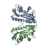
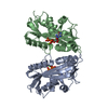
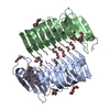
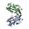
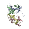
| Title | Crystal structure of human inosine triphosphate pyrophosphatase P32T variant | ||||||
 Components Components | Inosine triphosphate pyrophosphatase | ||||||
 Keywords Keywords | HYDROLASE | ||||||
| Function / homology |  Function and homology information Function and homology informationITP catabolic process / deoxyribonucleoside triphosphate catabolic process / XTP/dITP diphosphatase / ITP diphosphatase activity / XTP diphosphatase activity / dITP diphosphatase activity / nucleoside triphosphate catabolic process / nucleoside triphosphate diphosphatase activity / Ribavirin ADME / Purine catabolism ...ITP catabolic process / deoxyribonucleoside triphosphate catabolic process / XTP/dITP diphosphatase / ITP diphosphatase activity / XTP diphosphatase activity / dITP diphosphatase activity / nucleoside triphosphate catabolic process / nucleoside triphosphate diphosphatase activity / Ribavirin ADME / Purine catabolism / chromosome organization / nucleotide binding / nucleoplasm / metal ion binding / identical protein binding / cytoplasm / cytosol Similarity search - Function | ||||||
| Biological species |  Homo sapiens (human) Homo sapiens (human) | ||||||
| Method |  X-RAY DIFFRACTION / MOLECULAR REPLACEMENT / Resolution: 2.07 Å X-RAY DIFFRACTION / MOLECULAR REPLACEMENT / Resolution: 2.07 Å | ||||||
 Authors Authors | Simone, P.D. / Pavlov, Y.I. / Borgstahl, G.E.O. | ||||||
 Citation Citation | Journal: J.Struct.Biol. / Year: 2013 Title: The human ITPA polymorphic variant P32T is destabilized by the unpacking of the hydrophobic core. Authors: Simone, P.D. / Struble, L.R. / Kellezi, A. / Brown, C.A. / Grabow, C.E. / Khutsishvili, I. / Marky, L.A. / Pavlov, Y.I. / Borgstahl, G.E. | ||||||
| History |
|
- Structure visualization
| Structure viewer | Molecule: MolmilJmol/JSmol |
|---|
- Downloads & links
-Download
| PDBx/mmCIF format | 4f95.cif.gz | 52.9 KB | Display | PDBx/mmCIF format |
|---|---|---|---|---|
| PDB format | pdb4f95.ent.gz | 37 KB | Display | PDB format |
| PDBx/mmJSON format | 4f95.json.gz | Tree view | PDBx/mmJSON format | |
| Others |  Other downloads Other downloads |
-Validation report
| Arichive directory | https://data.pdbj.org/pub/pdb/validation_reports/f9/4f95ftp://data.pdbj.org/pub/pdb/validation_reports/f9/4f95 | HTTPS FTP |
|---|
-Related structure data
| Related structure data |  2i5dS S: Starting model for refinement |
|---|---|
| Similar structure data |


-Links
 PDBj
PDBj- Assembly
| Deposited unit | 
| |||||||||
|---|---|---|---|---|---|---|---|---|---|---|
| 1 | 
| |||||||||
| Unit cell |
| |||||||||
| Components on special symmetry positions |
|
-Components
| #1: Protein | Mass: 21754.893 Da / Num. of mol.: 1 / Mutation: P32T Source method: isolated from a genetically manipulated source Source: (gene. exp.) Homo sapiens (human) / Gene: C20orf37, ITPA, ITPA (P32T MUTANT), My049, OK/SW-cl.9 / Plasmid: PET-15B / Production host:  References: UniProt: Q9BY32, nucleoside-triphosphate diphosphatase |
|---|---|
| #2: Water | ChemComp-HOH /  Mass: 18.015 Da / Num. of mol.: 68 / Source method: isolated from a natural source / Formula: H2O Mass: 18.015 Da / Num. of mol.: 68 / Source method: isolated from a natural source / Formula: H2O |
-Experimental details
-Experiment
| Experiment | Method: X-RAY DIFFRACTION / Number of used crystals: 1 |
|---|
- Sample preparation
| Crystal | Density Matthews: 1.87 Å3/Da / Density % sol: 34.38 % |
|---|---|
| Crystal grow | Temperature: 295.15 K / Method: vapor diffusion, hanging drop / pH: 6.667 Details: 24.4% PEG 3350, 0.1 M BIS-TRIS, 10 MM BETA-MERCAPTOETHANOL, pH 6.667, VAPOR DIFFUSION, HANGING DROP, temperature 295.15K |
-Data collection
| Diffraction | Mean temperature: 100 K |
|---|---|
| Diffraction source | Source: ROTATING ANODE / Type: RIGAKU FR-E SUPERBRIGHT / Wavelength: 1.54 / Wavelength: 1.54 Å |
| Detector | Type: RIGAKU RAXIS IV++ / Detector: IMAGE PLATE / Date: Jul 29, 2010 |
| Radiation | Monochromator: OSMIC VARIMAX CONFOCAL MAX-FLUX OPTIC / Protocol: SINGLE WAVELENGTH / Monochromatic (M) / Laue (L): M / Scattering type: x-ray |
| Radiation wavelength | Wavelength: 1.54 Å / Relative weight: 1 |
| Reflection | Resolution: 2.07→29.74 Å / Num. all: 10531 / Num. obs: 10531 / % possible obs: 100 % / Observed criterion σ(F): 3 / Observed criterion σ(I): 3 / Redundancy: 3.66 % / Biso Wilson estimate: 44.886 Å2 / Rmerge(I) obs: 0.057 / Net I/σ(I): 9.9 |
| Reflection shell | Resolution: 2.07→2.14 Å / Redundancy: 3.71 % / Rmerge(I) obs: 0.478 / Mean I/σ(I) obs: 2.1 / % possible all: 100 |
- Processing
| Software |
| |||||||||||||||||||||||||||||||||||
|---|---|---|---|---|---|---|---|---|---|---|---|---|---|---|---|---|---|---|---|---|---|---|---|---|---|---|---|---|---|---|---|---|---|---|---|---|
| Refinement | Method to determine structure: MOLECULAR REPLACEMENT Starting model: PDB ENTRY 2I5D Resolution: 2.07→29.736 Å / SU ML: 0.37 / Isotropic thermal model: Isotropic / σ(F): 1.34 / Phase error: 32.74 / Stereochemistry target values: ML
| |||||||||||||||||||||||||||||||||||
| Solvent computation | Shrinkage radii: 0.86 Å / VDW probe radii: 1.1 Å / Solvent model: FLAT BULK SOLVENT MODEL / Bsol: 42.9 Å2 / ksol: 0.33 e/Å3 | |||||||||||||||||||||||||||||||||||
| Displacement parameters | Biso mean: 49 Å2
| |||||||||||||||||||||||||||||||||||
| Refinement step | Cycle: LAST / Resolution: 2.07→29.736 Å
| |||||||||||||||||||||||||||||||||||
| Refine LS restraints |
| |||||||||||||||||||||||||||||||||||
| LS refinement shell |
|
