 Movie
Movie Controller

[English] 日本語
 Yorodumi
- PDB-1vp2: CRYSTAL STRUCTURE OF A PUTATIVE XANTHOSINE TRIPHOSPHATE PYROPHOSP... -
+ Open data

- Basic information
| Entry | Database: PDB / ID: 1vp2 | ||||||
|---|---|---|---|---|---|---|---|
| Title | CRYSTAL STRUCTURE OF A PUTATIVE XANTHOSINE TRIPHOSPHATE PYROPHOSPHATASE/HAM1 PROTEIN HOMOLOG (TM0159) FROM THERMOTOGA MARITIMA AT 1.78 A RESOLUTION | ||||||
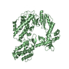
 Components Components | Putative Xanthosine triphosphate pyrophosphatase/HAM1 protein homolog | ||||||
 Keywords Keywords | HYDROLASE / PUTATIVE XANTHOSINE TRIPHOSPHATE PYROPHOSPHATASE / STRUCTURAL GENOMICS / JOINT CENTER FOR STRUCTURAL GENOMICS / JCSG / PROTEIN STRUCTURE INITIATIVE / PSI | ||||||
| Function / homology |  Function and homology information Function and homology informationpurine nucleoside triphosphate catabolic process / XTP/dITP diphosphatase / ITP diphosphatase activity / XTP diphosphatase activity / dITP diphosphatase activity / nucleoside triphosphate catabolic process / nucleoside triphosphate diphosphatase activity / nucleotide metabolic process / ribonucleoside triphosphate phosphatase activity / nucleotide binding ...purine nucleoside triphosphate catabolic process / XTP/dITP diphosphatase / ITP diphosphatase activity / XTP diphosphatase activity / dITP diphosphatase activity / nucleoside triphosphate catabolic process / nucleoside triphosphate diphosphatase activity / nucleotide metabolic process / ribonucleoside triphosphate phosphatase activity / nucleotide binding / metal ion binding / cytoplasm / cytosol Similarity search - Function | ||||||
| Biological species |   Thermotoga maritima (bacteria) Thermotoga maritima (bacteria) | ||||||
| Method |  X-RAY DIFFRACTION / SYNCHROTRON / MOLECULAR REPLACEMENT / Resolution: 1.78 Å X-RAY DIFFRACTION / SYNCHROTRON / MOLECULAR REPLACEMENT / Resolution: 1.78 Å | ||||||
 Authors Authors | Joint Center for Structural Genomics (JCSG) | ||||||
 Citation Citation | Journal: To be published Title: Crystal structure of Putative Xanthosine triphosphate pyrophosphatase1/HAM1 protein homolog (TM0159) from Thermotoga maritima at 1.78 A resolution Authors: Joint Center for Structural Genomics (JCSG) | ||||||
| History |
| ||||||
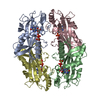
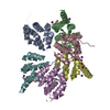
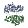
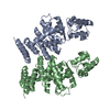
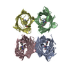
| Remark 300 | BIOMOLECULE: 1 THIS ENTRY CONTAINS THE CRYSTALLOGRAPHIC ASYMMETRIC UNIT WHICH CONSISTS OF 2 ... BIOMOLECULE: 1 THIS ENTRY CONTAINS THE CRYSTALLOGRAPHIC ASYMMETRIC UNIT WHICH CONSISTS OF 2 CHAIN(S). SEE REMARK 350 FOR INFORMATION ON GENERATING THE BIOLOGICAL MOLECULE(S). SIZE EXCLUSION CHROMATOGRAPHY DATA SUPPORT THE ASSIGNMENT OF THE TETRAMER AS THE BIOLOGICALLY SIGNIFICANT STATE. |
- Structure visualization
| Structure viewer | Molecule: MolmilJmol/JSmol |
|---|
- Downloads & links
-Download
| PDBx/mmCIF format | 1vp2.cif.gz | 93.2 KB | Display | PDBx/mmCIF format |
|---|---|---|---|---|
| PDB format | pdb1vp2.ent.gz | 70.5 KB | Display | PDB format |
| PDBx/mmJSON format | 1vp2.json.gz | Tree view | PDBx/mmJSON format | |
| Others |  Other downloads Other downloads |
-Validation report
| Arichive directory | https://data.pdbj.org/pub/pdb/validation_reports/vp/1vp2ftp://data.pdbj.org/pub/pdb/validation_reports/vp/1vp2 | HTTPS FTP |
|---|
-Related structure data
| Related structure data | 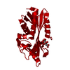 1v7rS S: Starting model for refinement |
|---|---|
| Similar structure data | |
| Other databases |
-Links
 PDBj
PDBj- Assembly
| Deposited unit | 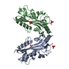
| ||||||||
|---|---|---|---|---|---|---|---|---|---|
| 1 | 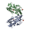
| ||||||||
| Unit cell |
|
-Components
| #1: Protein | Mass: 23834.539 Da / Num. of mol.: 2 Source method: isolated from a genetically manipulated source Source: (gene. exp.) Thermotoga maritima (bacteria) / Gene: TM0159 / Production host: References: UniProt: Q9WY06, Hydrolases; Acting on acid anhydrides; In phosphorus-containing anhydrides #2: Chemical | ChemComp-SO4 /   Mass: 96.063 Da / Num. of mol.: 4 / Source method: obtained synthetically / Formula: SO4 Mass: 96.063 Da / Num. of mol.: 4 / Source method: obtained synthetically / Formula: SO4#3: Water | ChemComp-HOH / |  Mass: 18.015 Da / Num. of mol.: 256 / Source method: isolated from a natural source / Formula: H2O Mass: 18.015 Da / Num. of mol.: 256 / Source method: isolated from a natural source / Formula: H2O |
|---|
-Experimental details
-Experiment
| Experiment | Method: X-RAY DIFFRACTION / Number of used crystals: 1 |
|---|
- Sample preparation
| Crystal | Density Matthews: 2.53 Å3/Da / Density % sol: 51 % |
|---|---|
| Crystal grow | Temperature: 277 K / Method: vapor diffusion, sitting drop, nanodrop Details: 1.6M (NH4)2SO4, 0.1M NaCl, 0.1M HEPES pH 7.5, VAPOR DIFFUSION,SITTING DROP,NANODROP, temperature 277K |
-Data collection
| Diffraction | Mean temperature: 100 K |
|---|---|
| Diffraction source | Source: SYNCHROTRON / Site: ALS  / Beamline: 5.0.1 / Wavelength: 1 / Beamline: 5.0.1 / Wavelength: 1 |
| Detector | Type: ADSC / Detector: CCD / Date: Mar 3, 2004 |
| Radiation | Monochromator: Single crystal, cylindrically bent, Si(220) / Protocol: SINGLE WAVELENGTH / Monochromatic (M) / Laue (L): M / Scattering type: x-ray |
| Radiation wavelength | Wavelength: 1 Å / Relative weight: 1 |
| Reflection | Resolution: 1.78→34.65 Å / Num. obs: 37599 / % possible obs: 83.6 % / Redundancy: 4.4 % / Biso Wilson estimate: 28.82 Å2 / Rsym value: 0.089 / Net I/σ(I): 12.6 |
| Reflection shell | Resolution: 1.78→1.83 Å / Redundancy: 3 % / Mean I/σ(I) obs: 1.8 / Num. unique all: 1359 / Rsym value: 0.502 / % possible all: 42.4 |
- Processing
| Software |
| ||||||||||||||||||||||||||||||||||||||||||||||||||||||||||||||||||||||||||||||||||||||||||||||||||||||||||||||||||||||||
|---|---|---|---|---|---|---|---|---|---|---|---|---|---|---|---|---|---|---|---|---|---|---|---|---|---|---|---|---|---|---|---|---|---|---|---|---|---|---|---|---|---|---|---|---|---|---|---|---|---|---|---|---|---|---|---|---|---|---|---|---|---|---|---|---|---|---|---|---|---|---|---|---|---|---|---|---|---|---|---|---|---|---|---|---|---|---|---|---|---|---|---|---|---|---|---|---|---|---|---|---|---|---|---|---|---|---|---|---|---|---|---|---|---|---|---|---|---|---|---|---|---|
| Refinement | Method to determine structure: MOLECULAR REPLACEMENT Starting model: 1V7R Resolution: 1.78→34.65 Å / Cor.coef. Fo:Fc: 0.962 / Cor.coef. Fo:Fc free: 0.941 / SU B: 4.286 / SU ML: 0.069 / TLS residual ADP flag: LIKELY RESIDUAL / Cross valid method: THROUGHOUT / ESU R: 0.121 / ESU R Free: 0.119 / Stereochemistry target values: MAXIMUM LIKELIHOOD / Details: HYDROGENS HAVE BEEN ADDED IN THE RIDING POSITIONS
| ||||||||||||||||||||||||||||||||||||||||||||||||||||||||||||||||||||||||||||||||||||||||||||||||||||||||||||||||||||||||
| Solvent computation | Ion probe radii: 0.8 Å / Shrinkage radii: 0.8 Å / VDW probe radii: 1.2 Å / Solvent model: BABINET MODEL WITH MASK | ||||||||||||||||||||||||||||||||||||||||||||||||||||||||||||||||||||||||||||||||||||||||||||||||||||||||||||||||||||||||
| Displacement parameters | Biso mean: 21.043 Å2
| ||||||||||||||||||||||||||||||||||||||||||||||||||||||||||||||||||||||||||||||||||||||||||||||||||||||||||||||||||||||||
| Refinement step | Cycle: LAST / Resolution: 1.78→34.65 Å
| ||||||||||||||||||||||||||||||||||||||||||||||||||||||||||||||||||||||||||||||||||||||||||||||||||||||||||||||||||||||||
| Refine LS restraints |
| ||||||||||||||||||||||||||||||||||||||||||||||||||||||||||||||||||||||||||||||||||||||||||||||||||||||||||||||||||||||||
| LS refinement shell | Resolution: 1.78→1.826 Å / Total num. of bins used: 20
| ||||||||||||||||||||||||||||||||||||||||||||||||||||||||||||||||||||||||||||||||||||||||||||||||||||||||||||||||||||||||
| Refinement TLS params. | Method: refined / Refine-ID: X-RAY DIFFRACTION
| ||||||||||||||||||||||||||||||||||||||||||||||||||||||||||||||||||||||||||||||||||||||||||||||||||||||||||||||||||||||||
| Refinement TLS group | Refine-ID: X-RAY DIFFRACTION / Selection: ALL / Auth seq-ID: 3 - 191 / Label seq-ID: 15 - 203
|
