Mass: 18.015 Da / Num. of mol.: 148 / Source method: isolated from a natural source / Formula: H2O
-
Experimental details
-
Experiment
Experiment
Method: X-RAY DIFFRACTION / Number of used crystals: 1
-
Sample preparation
Crystal
Density Matthews: 2.39 Å3/Da / Density % sol: 48.48 %
Crystal grow
Temperature: 298 K / Method: vapor diffusion, sitting drop / pH: 5.4 Details: Protein crystallized from 2.5 mM Ammonium Sulfate and 0.1M sodium Citrate, pH 5.4, VAPOR DIFFUSION, SITTING DROP, temperature 298K
-
Data collection
Diffraction
Mean temperature: 100 K
Diffraction source
Source: ROTATING ANODE / Type: RIGAKU / Wavelength: 1.5412 Å
Detector
Type: RIGAKU RAXIS IV++ / Detector: IMAGE PLATE
Radiation
Protocol: SINGLE WAVELENGTH / Monochromatic (M) / Laue (L): M / Scattering type: x-ray
Radiation wavelength
Wavelength: 1.5412 Å / Relative weight: 1
Reflection
Resolution: 1.64→50 Å / Num. obs: 13828 / % possible obs: 98 % / Redundancy: 3.3 % / Rmerge(I) obs: 0.049 / Χ2: 1.343 / Net I/σ(I): 49.9
Reflection shell
Resolution (Å)
Redundancy (%)
Rmerge(I) obs
Num. unique all
Χ2
Diffraction-ID
% possible all
1.64-1.67
3.4
0.112
676
1.992
1
98.1
1.67-1.7
3.5
0.101
679
1.919
1
98.3
1.7-1.73
3.5
0.087
672
1.807
1
98.4
1.73-1.77
3.5
0.086
655
1.734
1
97.9
1.77-1.81
3.5
0.083
694
1.833
1
97.5
1.81-1.85
3.4
0.076
680
1.652
1
97.3
1.85-1.89
3.4
0.071
669
1.637
1
97
1.89-1.94
3.3
0.064
667
1.619
1
97.4
1.94-2
3.3
0.06
667
1.405
1
98.2
2-2.07
3.3
0.056
700
1.41
1
97.8
2.07-2.14
3.2
0.053
683
1.225
1
98.6
2.14-2.23
3.2
0.051
680
1.223
1
98.1
2.23-2.33
3.2
0.048
691
1.129
1
97.6
2.33-2.45
3.2
0.051
708
1.14
1
99.4
2.45-2.6
3.2
0.051
685
1.064
1
98.7
2.6-2.8
3.3
0.049
696
0.957
1
98.4
2.8-3.09
3.3
0.049
716
0.9
1
98.5
3.09-3.53
3.3
0.046
713
0.774
1
98.3
3.53-4.45
3.4
0.045
718
0.706
1
98.6
4.45-50
3.3
0.048
779
0.737
1
96.5
-
Processing
Software
Name
Version
Classification
NB
d*TREK
datascaling
SCALEPACK
datascaling
PHENIX
1.7.3_928
refinement
PDB_EXTRACT
3.11
dataextraction
d*TREK
datareduction
Refinement
Method to determine structure: FOURIER SYNTHESIS / Resolution: 1.64→24.545 Å / Occupancy max: 1 / Occupancy min: 0 / FOM work R set: 0.8827 / SU ML: 0.23 / σ(F): 1.34 / Phase error: 17.73 / Stereochemistry target values: ML
Rfactor
Num. reflection
% reflection
Rfree
0.2019
1379
10 %
Rwork
0.1801
-
-
obs
0.1823
13789
97.95 %
Solvent computation
Shrinkage radii: 0.86 Å / VDW probe radii: 1.1 Å / Solvent model: FLAT BULK SOLVENT MODEL / Bsol: 34.958 Å2 / ksol: 0.381 e/Å3
In the structure databanks used in Yorodumi, some data are registered as the other names, "COVID-19 virus" and "2019-nCoV". Here are the details of the virus and the list of structure data.
Jan 31, 2019. EMDB accession codes are about to change! (news from PDBe EMDB page)
EMDB accession codes are about to change! (news from PDBe EMDB page)
The allocation of 4 digits for EMDB accession codes will soon come to an end. Whilst these codes will remain in use, new EMDB accession codes will include an additional digit and will expand incrementally as the available range of codes is exhausted. The current 4-digit format prefixed with “EMD-” (i.e. EMD-XXXX) will advance to a 5-digit format (i.e. EMD-XXXXX), and so on. It is currently estimated that the 4-digit codes will be depleted around Spring 2019, at which point the 5-digit format will come into force.
The EM Navigator/Yorodumi systems omit the EMD- prefix.
Related info.:Q: What is EMD? / ID/Accession-code notation in Yorodumi/EM Navigator
Yorodumi is a browser for structure data from EMDB, PDB, SASBDB, etc.
This page is also the successor to EM Navigator detail page, and also detail information page/front-end page for Omokage search.
The word "yorodu" (or yorozu) is an old Japanese word meaning "ten thousand". "mi" (miru) is to see.
Related info.:EMDB / PDB / SASBDB / Comparison of 3 databanks / Yorodumi Search / Aug 31, 2016. New EM Navigator & Yorodumi / Yorodumi Papers / Jmol/JSmol / Function and homology information / Changes in new EM Navigator and Yorodumi
 Movie
Movie Controller

 Yorodumi
 Open data
 Basic information
 Components
 Keywords
 Function and homology information
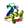
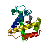
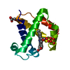
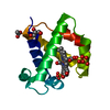
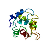
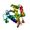
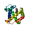
 Cerebratulus lacteus (milky ribbon-worm)
 X-RAY DIFFRACTION /
  Authors
 Citation
 Structure visualization
 Downloads & links
 Other downloads


 PDBj
PDBj


 Assembly


 Mass: 616.487 Da / Num. of mol.: 1 / Source method: obtained synthetically / Formula: C34H32FeN4O4
 Mass: 96.063 Da / Num. of mol.: 1 / Source method: obtained synthetically / Formula: SO4
 Mass: 92.094 Da / Num. of mol.: 1 / Source method: obtained synthetically / Formula: C3H8O3
 Mass: 18.015 Da / Num. of mol.: 148 / Source method: isolated from a natural source / Formula: H2O
 Sample preparation
 Processing