 Movie
Movie Controller

[English] 日本語
 Yorodumi
- PDB-1kr7: Crystal structure of the nerve tissue mini-hemoglobin from the ne... -
+ Open data

- Basic information
| Entry | Database: PDB / ID: 1kr7 | ||||||
|---|---|---|---|---|---|---|---|
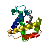
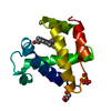
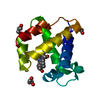
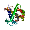
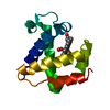
| Title | Crystal structure of the nerve tissue mini-hemoglobin from the nemertean worm Cerebratulus lacteus | ||||||
 Components Components | Neural globin | ||||||
 Keywords Keywords | OXYGEN STORAGE/TRANSPORT / nerve tissue / mini-hemoglobin / protein cavities / Oxygen transport / OXYGEN STORAGE-TRANSPORT COMPLEX | ||||||
| Function / homology |  Function and homology information Function and homology informationoxygen carrier activity / oxygen binding / heme binding / metal ion binding Similarity search - Function | ||||||
| Biological species |  Cerebratulus lacteus (milky ribbon-worm) Cerebratulus lacteus (milky ribbon-worm) | ||||||
| Method |  X-RAY DIFFRACTION / SYNCHROTRON / MAD / Resolution: 1.5 Å X-RAY DIFFRACTION / SYNCHROTRON / MAD / Resolution: 1.5 Å | ||||||
 Authors Authors | Pesce, A. / Nardini, M. / Dewilde, S. / Geuens, E. / Yamauchi, k. / Ascenzi, P. / Riggs, A.F. / Moens, L. / Bolognesi, M. | ||||||
 Citation Citation | Journal: Structure / Year: 2002 Title: The 109 residue nerve tissue minihemoglobin from Cerebratulus lacteus highlights striking structural plasticity of the alpha-helical globin fold Authors: Pesce, A. / Nardini, M. / Dewilde, S. / Geuens, E. / Yamauchi, k. / Ascenzi, P. / Riggs, A.F. / Moens, L. / Bolognesi, M. #1: Journal: Acta Crystallogr.,Sect.D / Year: 2001Title: Crystallization and preliminary X-ray analysis of neural hemoglobin from the nemertean worm Cerebratulus lacteus Authors: Pesce, A. / Nardini, M. / Dewilde, S. / Ascenzi, P. / Riggs, A.F. / Yamauchi, K. / Geuens, E. / Moens, L. / Bolognesi, M. | ||||||
| History |
|
- Structure visualization
| Structure viewer | Molecule: MolmilJmol/JSmol |
|---|
- Downloads & links
-Download
| PDBx/mmCIF format | 1kr7.cif.gz | 60.8 KB | Display | PDBx/mmCIF format |
|---|---|---|---|---|
| PDB format | pdb1kr7.ent.gz | 44.1 KB | Display | PDB format |
| PDBx/mmJSON format | 1kr7.json.gz | Tree view | PDBx/mmJSON format | |
| Others |  Other downloads Other downloads |
-Validation report
| Arichive directory | https://data.pdbj.org/pub/pdb/validation_reports/kr/1kr7ftp://data.pdbj.org/pub/pdb/validation_reports/kr/1kr7 | HTTPS FTP |
|---|
-Related structure data
| Similar structure data |
|---|


-Links
 PDBj
PDBj


- Assembly
| Deposited unit | 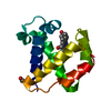
| ||||||||
|---|---|---|---|---|---|---|---|---|---|
| 1 |
| ||||||||
| Unit cell |
|
-Components
-Protein , 1 types, 1 molecules A
| #1: Protein | Mass: 11546.949 Da / Num. of mol.: 1 Source method: isolated from a genetically manipulated source Source: (gene. exp.) Cerebratulus lacteus (milky ribbon-worm)Plasmid: pET3a / Production host:  |
|---|
-Non-polymers , 5 types, 110 molecules 


| #2: Chemical | ChemComp-SO4 /  Mass: 96.063 Da / Num. of mol.: 1 / Source method: obtained synthetically / Formula: SO4 Mass: 96.063 Da / Num. of mol.: 1 / Source method: obtained synthetically / Formula: SO4 |
|---|---|
| #3: Chemical | ChemComp-ACT /  Mass: 59.044 Da / Num. of mol.: 1 / Source method: obtained synthetically / Formula: C2H3O2 Mass: 59.044 Da / Num. of mol.: 1 / Source method: obtained synthetically / Formula: C2H3O2 |
| #4: Chemical | ChemComp-HEM /  Mass: 616.487 Da / Num. of mol.: 1 / Source method: obtained synthetically / Formula: C34H32FeN4O4 Mass: 616.487 Da / Num. of mol.: 1 / Source method: obtained synthetically / Formula: C34H32FeN4O4 |
| #5: Chemical | ChemComp-OXY /  Mass: 31.999 Da / Num. of mol.: 1 / Source method: obtained synthetically / Formula: O2 Mass: 31.999 Da / Num. of mol.: 1 / Source method: obtained synthetically / Formula: O2 |
| #6: Water | ChemComp-HOH / Mass: 18.015 Da / Num. of mol.: 106 / Source method: isolated from a natural source / Formula: H2O |
-Experimental details
-Experiment
| Experiment | Method: X-RAY DIFFRACTION / Number of used crystals: 1 |
|---|
- Sample preparation
| Crystal | Density Matthews: 2.3 Å3/Da / Density % sol: 46.5 % | ||||||||||||||||||||||||
|---|---|---|---|---|---|---|---|---|---|---|---|---|---|---|---|---|---|---|---|---|---|---|---|---|---|
| Crystal grow | Temperature: 277 K / Method: vapor diffusion, hanging drop / pH: 5.5 Details: ammonium sulfate, sodium acetate, pH 5.5, VAPOR DIFFUSION, HANGING DROP, temperature 277K | ||||||||||||||||||||||||
| Crystal grow | *PLUS Temperature: 4 ℃ / Method: vapor diffusion | ||||||||||||||||||||||||
| Components of the solutions | *PLUS
|
-Data collection
| Diffraction | Mean temperature: 100 K | ||||||||||||
|---|---|---|---|---|---|---|---|---|---|---|---|---|---|
| Diffraction source | Source: SYNCHROTRON / Site: ESRF  / Beamline: ID29 / Wavelength: 1.739, 1.740, 0.915 / Beamline: ID29 / Wavelength: 1.739, 1.740, 0.915 | ||||||||||||
| Detector | Type: ADSC QUANTUM 4 / Detector: CCD / Date: Jun 23, 2001 / Details: mirrors | ||||||||||||
| Radiation | Monochromator: Si 311 channel and Si 111 channel cut / Protocol: MAD / Monochromatic (M) / Laue (L): M / Scattering type: x-ray | ||||||||||||
| Radiation wavelength |
| ||||||||||||
| Reflection | Resolution: 1.5→35 Å / Num. all: 17906 / Num. obs: 17906 / % possible obs: 97.1 % / Observed criterion σ(F): 0 / Observed criterion σ(I): 0 / Redundancy: 3.8 % / Biso Wilson estimate: 14 Å2 / Rmerge(I) obs: 0.054 / Rsym value: 0.054 / Net I/σ(I): 14.3 | ||||||||||||
| Reflection shell | Resolution: 1.5→1.53 Å / Redundancy: 3.8 % / Rmerge(I) obs: 0.239 / Mean I/σ(I) obs: 3.2 / Num. unique all: 1168 / Rsym value: 0.239 / % possible all: 95.7 | ||||||||||||
| Reflection | *PLUS Highest resolution: 1.5 Å / Lowest resolution: 35 Å / Num. measured all: 67606 / Rmerge(I) obs: 0.054 | ||||||||||||
| Reflection shell | *PLUS % possible obs: 95.7 % / Rmerge(I) obs: 0.239 |
- Processing
| Software |
| |||||||||||||||||||||||||
|---|---|---|---|---|---|---|---|---|---|---|---|---|---|---|---|---|---|---|---|---|---|---|---|---|---|---|
| Refinement | Method to determine structure: MAD / Resolution: 1.5→35.14 Å / SU B: 2.82431 / SU ML: 0.04376 / Cross valid method: THROUGHOUT / σ(F): 0 / σ(I): 0 / ESU R: 0.08727 / ESU R Free: 0.07211 / Stereochemistry target values: Engh & Huber
| |||||||||||||||||||||||||
| Solvent computation | Ion probe radii: 0.8 Å / Shrinkage radii: 0.8 Å / VDW probe radii: 1.4 Å | |||||||||||||||||||||||||
| Displacement parameters | Biso mean: 16.023 Å2
| |||||||||||||||||||||||||
| Refinement step | Cycle: LAST / Resolution: 1.5→35.14 Å
| |||||||||||||||||||||||||
| Refine LS restraints |
| |||||||||||||||||||||||||
| Refinement | *PLUS Highest resolution: 1.5 Å / Lowest resolution: 35 Å / Rfactor all: 0.1563 / Rfactor obs: 0.153 / Rfactor Rfree: 0.18702 / Rfactor Rwork: 0.15298 | |||||||||||||||||||||||||
| Solvent computation | *PLUS | |||||||||||||||||||||||||
| Displacement parameters | *PLUS | |||||||||||||||||||||||||
| Refine LS restraints | *PLUS
|
