 Movie
Movie Controller


+ Open data

- Basic information
| Entry | Database: PDB / ID: 4f1y | ||||||
|---|---|---|---|---|---|---|---|
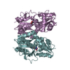
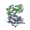
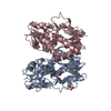
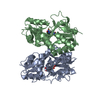
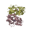
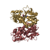
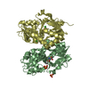
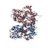
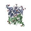
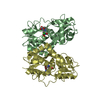
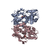
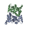
| Title | CNQX bound to the ligand binding domain of GluA3 | ||||||
 Components Components | Glutamate receptor 3 | ||||||
 Keywords Keywords | TRANSPORT PROTEIN/AGONIST / glutamate receptor / GluA3 / GluR3 / AMPA receptor / S1S2 / LBD / neurotransmitter receptor / CNQX / TRANSPORT PROTEIN / TRANSPORT PROTEIN-AGONIST complex | ||||||
| Function / homology |  Function and homology information Function and homology informationTrafficking of AMPA receptors / ligand-gated calcium channel activity / Synaptic adhesion-like molecules / Activation of AMPA receptors / protein heterotetramerization / Trafficking of GluR2-containing AMPA receptors / response to lithium ion / AMPA glutamate receptor activity / parallel fiber to Purkinje cell synapse / regulation of receptor recycling ...Trafficking of AMPA receptors / ligand-gated calcium channel activity / Synaptic adhesion-like molecules / Activation of AMPA receptors / protein heterotetramerization / Trafficking of GluR2-containing AMPA receptors / response to lithium ion / AMPA glutamate receptor activity / parallel fiber to Purkinje cell synapse / regulation of receptor recycling / AMPA glutamate receptor complex / cellular response to glycine / ionotropic glutamate receptor complex / asymmetric synapse / Unblocking of NMDA receptors, glutamate binding and activation / synaptic cleft / response to fungicide / glutamate-gated receptor activity / glutamate-gated calcium ion channel activity / presynaptic active zone membrane / ligand-gated monoatomic ion channel activity involved in regulation of presynaptic membrane potential / dendritic shaft / synaptic transmission, glutamatergic / transmitter-gated monoatomic ion channel activity involved in regulation of postsynaptic membrane potential / postsynaptic density membrane / modulation of chemical synaptic transmission / long-term synaptic potentiation / terminal bouton / amyloid-beta binding / presynaptic membrane / protein homotetramerization / dendritic spine / perikaryon / postsynaptic membrane / postsynaptic density / neuronal cell body / dendrite / glutamatergic synapse / protein-containing complex / membrane / plasma membrane Similarity search - Function | ||||||
| Biological species |  | ||||||
| Method |  X-RAY DIFFRACTION / SYNCHROTRON / MOLECULAR REPLACEMENT / Resolution: 1.79 Å X-RAY DIFFRACTION / SYNCHROTRON / MOLECULAR REPLACEMENT / Resolution: 1.79 Å | ||||||
 Authors Authors | Ahmed, A.H. / Oswald, R.E. | ||||||
 Citation Citation | Journal: Biochemistry / Year: 2012 Title: The loss of an electrostatic contact unique to AMPA receptor ligand binding domain 2 slows channel activation. Authors: Holley, S.M. / Ahmed, A.H. / Srinivasan, J. / Murthy, S.E. / Weiland, G.A. / Oswald, R.E. / Nowak, L.M. | ||||||
| History |
|
- Structure visualization
| Structure viewer | Molecule: MolmilJmol/JSmol |
|---|
- Downloads & links
-Download
| PDBx/mmCIF format | 4f1y.cif.gz | 125.3 KB | Display | PDBx/mmCIF format |
|---|---|---|---|---|
| PDB format | pdb4f1y.ent.gz | 95.7 KB | Display | PDB format |
| PDBx/mmJSON format | 4f1y.json.gz | Tree view | PDBx/mmJSON format | |
| Others |  Other downloads Other downloads |
-Validation report
| Arichive directory | https://data.pdbj.org/pub/pdb/validation_reports/f1/4f1yftp://data.pdbj.org/pub/pdb/validation_reports/f1/4f1y | HTTPS FTP |
|---|
-Related structure data
| Related structure data |  4f22C 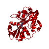 4f29C 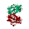 4f2oC 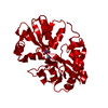 4f2qC  4f31C 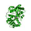 4f39C 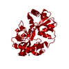 4f3bC  4f3gC  3b7dS C: citing same article ( S: Starting model for refinement |
|---|---|
| Similar structure data |
-Links
 PDBj
PDBj


- Assembly
| Deposited unit | 
| ||||||||
|---|---|---|---|---|---|---|---|---|---|
| 1 |
| ||||||||
| Unit cell |
|
-Components
| #1: Protein | Mass: 28970.402 Da / Num. of mol.: 2 Source method: isolated from a genetically manipulated source Source: (gene. exp.)  #2: Chemical |   Mass: 230.137 Da / Num. of mol.: 2 / Source method: obtained synthetically / Formula: C9H2N4O4 Mass: 230.137 Da / Num. of mol.: 2 / Source method: obtained synthetically / Formula: C9H2N4O4#3: Chemical |   Mass: 65.409 Da / Num. of mol.: 2 / Source method: obtained synthetically / Formula: Zn Mass: 65.409 Da / Num. of mol.: 2 / Source method: obtained synthetically / Formula: Zn#4: Water | ChemComp-HOH / |  Mass: 18.015 Da / Num. of mol.: 492 / Source method: isolated from a natural source / Formula: H2O Mass: 18.015 Da / Num. of mol.: 492 / Source method: isolated from a natural source / Formula: H2OHas protein modification | Y | |
|---|
-Experimental details
-Experiment
| Experiment | Method: X-RAY DIFFRACTION / Number of used crystals: 1 |
|---|
- Sample preparation
| Crystal | Density Matthews: 2.49 Å3/Da / Density % sol: 50.68 % |
|---|---|
| Crystal grow | Temperature: 277 K / Method: vapor diffusion, hanging drop / pH: 6.5 Details: 14-15% PEG 8K, 0.1 M sodium cacodylate, 0.1-0.15 M zinc acetate, 0.25 M ammonium sulfate , pH 6.5, VAPOR DIFFUSION, HANGING DROP, temperature 277K |
-Data collection
| Diffraction | Mean temperature: 100 K |
|---|---|
| Diffraction source | Source: SYNCHROTRON / Site: CHESS  / Beamline: A1 / Wavelength: 0.977 Å / Beamline: A1 / Wavelength: 0.977 Å |
| Detector | Type: ADSC QUANTUM 210 / Detector: CCD / Date: Mar 14, 2010 |
| Radiation | Monochromator: Rh coated Si / Protocol: SINGLE WAVELENGTH / Monochromatic (M) / Laue (L): M / Scattering type: x-ray |
| Radiation wavelength | Wavelength: 0.977 Å / Relative weight: 1 |
| Reflection | Resolution: 1.79→50 Å / % possible obs: 95.7 % / Observed criterion σ(F): 0 / Observed criterion σ(I): -3 / Redundancy: 2.9 % / Rmerge(I) obs: 0.087 / Rsym value: 0.087 / Net I/σ(I): 23.26 |
| Reflection shell | Resolution: 1.8→1.83 Å / Redundancy: 2.5 % / Rmerge(I) obs: 0.489 / Mean I/σ(I) obs: 2.059 / Rsym value: 0.489 / % possible all: 98.2 |
- Processing
| Software |
| ||||||||||||||||||||||||||||||||||||||||||||||||||||||||||||||||||||||||||||||||||||||||||||||||||
|---|---|---|---|---|---|---|---|---|---|---|---|---|---|---|---|---|---|---|---|---|---|---|---|---|---|---|---|---|---|---|---|---|---|---|---|---|---|---|---|---|---|---|---|---|---|---|---|---|---|---|---|---|---|---|---|---|---|---|---|---|---|---|---|---|---|---|---|---|---|---|---|---|---|---|---|---|---|---|---|---|---|---|---|---|---|---|---|---|---|---|---|---|---|---|---|---|---|---|---|
| Refinement | Method to determine structure: MOLECULAR REPLACEMENT Starting model: 3B7D Resolution: 1.79→26.779 Å / SU ML: 0.27 / Isotropic thermal model: Isotropic / Cross valid method: THROUGHOUT / σ(F): 0.05 / Phase error: 26.13 / Stereochemistry target values: ML
| ||||||||||||||||||||||||||||||||||||||||||||||||||||||||||||||||||||||||||||||||||||||||||||||||||
| Solvent computation | Shrinkage radii: 0.9 Å / VDW probe radii: 1.11 Å / Solvent model: FLAT BULK SOLVENT MODEL / Bsol: 50.983 Å2 / ksol: 0.378 e/Å3 | ||||||||||||||||||||||||||||||||||||||||||||||||||||||||||||||||||||||||||||||||||||||||||||||||||
| Displacement parameters |
| ||||||||||||||||||||||||||||||||||||||||||||||||||||||||||||||||||||||||||||||||||||||||||||||||||
| Refinement step | Cycle: LAST / Resolution: 1.79→26.779 Å
| ||||||||||||||||||||||||||||||||||||||||||||||||||||||||||||||||||||||||||||||||||||||||||||||||||
| Refine LS restraints |
| ||||||||||||||||||||||||||||||||||||||||||||||||||||||||||||||||||||||||||||||||||||||||||||||||||
| LS refinement shell |
|
