- PDB-4h8j: Structure of GluA2-LBD in complex with MES -
+
Open data
ID or keywords:
Loading...
-
Basic information
Entry
Database: PDB / ID: 4h8j
Title
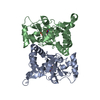
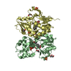
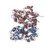
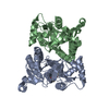
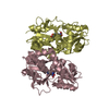
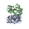
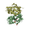
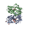
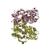
Structure of GluA2-LBD in complex with MES
Components
Glutamate receptor 2
Keywords
SIGNALING PROTEIN / membrane protein / glycoprotein / transmembrane protein / ligand-binding domain / ion transport / ion channel / ionotropic glutamate receptor / AMPA receptor / MES / synapse / postsynaptic cell membrane
Function / homology
Function and homology information
spine synapse / dendritic spine neck / dendritic spine cytoplasm / dendritic spine head / cellular response to amine stimulus / Activation of AMPA receptors / ligand-gated monoatomic cation channel activity / perisynaptic space / Trafficking of GluR2-containing AMPA receptors / response to lithium ion ...spine synapse / dendritic spine neck / dendritic spine cytoplasm / dendritic spine head / cellular response to amine stimulus / Activation of AMPA receptors / ligand-gated monoatomic cation channel activity / perisynaptic space / Trafficking of GluR2-containing AMPA receptors / response to lithium ion / AMPA glutamate receptor activity / AMPA glutamate receptor clustering / regulation of receptor recycling / kainate selective glutamate receptor activity / immunoglobulin binding / AMPA glutamate receptor complex / extracellularly glutamate-gated ion channel activity / cellular response to glycine / ionotropic glutamate receptor complex / asymmetric synapse / Unblocking of NMDA receptors, glutamate binding and activation / glutamate receptor binding / positive regulation of synaptic transmission / conditioned place preference / regulation of synaptic transmission, glutamatergic / response to fungicide / extracellular ligand-gated monoatomic ion channel activity / cytoskeletal protein binding / glutamate-gated receptor activity / cellular response to brain-derived neurotrophic factor stimulus / regulation of long-term synaptic depression / glutamate-gated calcium ion channel activity / somatodendritic compartment / presynaptic active zone membrane / ionotropic glutamate receptor signaling pathway / ionotropic glutamate receptor binding / dendrite cytoplasm / dendrite membrane / excitatory synapse / ligand-gated monoatomic ion channel activity involved in regulation of presynaptic membrane potential / positive regulation of excitatory postsynaptic potential / dendritic shaft / SNARE binding / synaptic membrane / PDZ domain binding / establishment of protein localization / protein tetramerization / synaptic transmission, glutamatergic / transmitter-gated monoatomic ion channel activity involved in regulation of postsynaptic membrane potential / receptor internalization / cerebral cortex development / postsynaptic density membrane / modulation of chemical synaptic transmission / long-term synaptic potentiation / Schaffer collateral - CA1 synapse / terminal bouton / synaptic vesicle / synaptic vesicle membrane / amyloid-beta binding / presynapse / growth cone / signaling receptor activity / presynaptic membrane / scaffold protein binding / chemical synaptic transmission / dendritic spine / perikaryon / postsynaptic membrane / postsynaptic density / neuron projection / external side of plasma membrane / axon / neuronal cell body / synapse / dendrite / protein kinase binding / protein-containing complex binding / glutamatergic synapse / cell surface / endoplasmic reticulum / protein-containing complex / membrane / identical protein binding / plasma membrane Similarity search - Function
Ionotropic glutamate receptor, metazoa / Ligated ion channel L-glutamate- and glycine-binding site / Ligand-gated ion channel / Ionotropic glutamate receptor, L-glutamate and glycine-binding domain / Ligated ion channel L-glutamate- and glycine-binding site / : / Ionotropic glutamate receptor / Eukaryotic homologues of bacterial periplasmic substrate binding proteins. / Periplasmic binding protein-like II / Receptor, ligand binding region ...Ionotropic glutamate receptor, metazoa / Ligated ion channel L-glutamate- and glycine-binding site / Ligand-gated ion channel / Ionotropic glutamate receptor, L-glutamate and glycine-binding domain / Ligated ion channel L-glutamate- and glycine-binding site / : / Ionotropic glutamate receptor / Eukaryotic homologues of bacterial periplasmic substrate binding proteins. / Periplasmic binding protein-like II / Receptor, ligand binding region / Receptor family ligand binding region / D-Maltodextrin-Binding Protein; domain 2 / Periplasmic binding protein-like I / 3-Layer(aba) Sandwich / Alpha Beta Similarity search - Domain/homology
In the structure databanks used in Yorodumi, some data are registered as the other names, "COVID-19 virus" and "2019-nCoV". Here are the details of the virus and the list of structure data.
Jan 31, 2019. EMDB accession codes are about to change! (news from PDBe EMDB page)
EMDB accession codes are about to change! (news from PDBe EMDB page)
The allocation of 4 digits for EMDB accession codes will soon come to an end. Whilst these codes will remain in use, new EMDB accession codes will include an additional digit and will expand incrementally as the available range of codes is exhausted. The current 4-digit format prefixed with “EMD-” (i.e. EMD-XXXX) will advance to a 5-digit format (i.e. EMD-XXXXX), and so on. It is currently estimated that the 4-digit codes will be depleted around Spring 2019, at which point the 5-digit format will come into force.
The EM Navigator/Yorodumi systems omit the EMD- prefix.
Related info.:Q: What is EMD? / ID/Accession-code notation in Yorodumi/EM Navigator
Yorodumi is a browser for structure data from EMDB, PDB, SASBDB, etc.
This page is also the successor to EM Navigator detail page, and also detail information page/front-end page for Omokage search.
The word "yorodu" (or yorozu) is an old Japanese word meaning "ten thousand". "mi" (miru) is to see.
Related info.:EMDB / PDB / SASBDB / Comparison of 3 databanks / Yorodumi Search / Aug 31, 2016. New EM Navigator & Yorodumi / Yorodumi Papers / Jmol/JSmol / Function and homology information / Changes in new EM Navigator and Yorodumi
 Movie
Movie Controller


 Open data
 Basic information
 Components
 Keywords
 Function and homology information
 X-RAY DIFFRACTION /
  Authors
 Citation
 Structure visualization
 Downloads & links
 Other downloads


 PDBj
PDBj


 Assembly
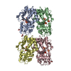


 Mass: 195.237 Da / Num. of mol.: 2 / Source method: obtained synthetically / Formula: C6H13NO4S / Comment: pH buffer*YM
 Mass: 96.063 Da / Num. of mol.: 12 / Source method: obtained synthetically / Formula: SO4
 Mass: 62.068 Da / Num. of mol.: 5 / Source method: obtained synthetically / Formula: C2H6O2
 Mass: 18.015 Da / Num. of mol.: 1089 / Source method: isolated from a natural source / Formula: H2O
 Sample preparation
 / Beamline: 14.1 / Wavelength: 0.91841 Å
 Processing