 Movie
Movie Controller


+ Open data

- Basic information
| Entry | Database: PDB / ID: 4f0g | ||||||
|---|---|---|---|---|---|---|---|
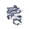
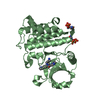
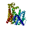
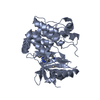
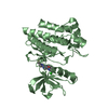
| Title | Crystal Structure of the Roco4 Kinase Domain from D. discoideum | ||||||
 Components Components | Serine/threonine-protein kinase roco4 | ||||||
 Keywords Keywords | TRANSFERASE / SIGNALING PROTEIN / Protein Kinase / LRRK2 / ROCO / ATP-binding / Nucleotide-binding / Parkinson disease / Serine/threonine-protein kinase | ||||||
| Function / homology |  Function and homology information Function and homology informationsorocarp stalk development / Regulation of necroptotic cell death / phototaxis / mitochondrial electron transport, NADH to ubiquinone / response to reactive oxygen species / protein tyrosine kinase activity / protein kinase activity / non-specific serine/threonine protein kinase / protein serine kinase activity / protein serine/threonine kinase activity ...sorocarp stalk development / Regulation of necroptotic cell death / phototaxis / mitochondrial electron transport, NADH to ubiquinone / response to reactive oxygen species / protein tyrosine kinase activity / protein kinase activity / non-specific serine/threonine protein kinase / protein serine kinase activity / protein serine/threonine kinase activity / GTP binding / signal transduction / mitochondrion / ATP binding / cytoplasm / cytosol Similarity search - Function | ||||||
| Biological species |  | ||||||
| Method |  X-RAY DIFFRACTION / SYNCHROTRON / MOLECULAR REPLACEMENT / Resolution: 2 Å X-RAY DIFFRACTION / SYNCHROTRON / MOLECULAR REPLACEMENT / Resolution: 2 Å | ||||||
 Authors Authors | Gilsbach, B.K. / Vetter, I.R. / Wittinghofer, A. / Kortholt, A. | ||||||
 Citation Citation | Journal: Proc.Natl.Acad.Sci.USA / Year: 2012 Title: Roco kinase structures give insights into the mechanism of Parkinson disease-related leucine-rich-repeat kinase 2 mutations. Authors: Gilsbach, B.K. / Ho, F.Y. / Vetter, I.R. / van Haastert, P.J. / Wittinghofer, A. / Kortholt, A. | ||||||
| History |
|
- Structure visualization
| Structure viewer | Molecule: MolmilJmol/JSmol |
|---|
- Downloads & links
-Download
| PDBx/mmCIF format | 4f0g.cif.gz | 68.2 KB | Display | PDBx/mmCIF format |
|---|---|---|---|---|
| PDB format | pdb4f0g.ent.gz | 49.1 KB | Display | PDB format |
| PDBx/mmJSON format | 4f0g.json.gz | Tree view | PDBx/mmJSON format | |
| Others |  Other downloads Other downloads |
-Validation report
| Arichive directory | https://data.pdbj.org/pub/pdb/validation_reports/f0/4f0gftp://data.pdbj.org/pub/pdb/validation_reports/f0/4f0g | HTTPS FTP |
|---|
-Related structure data
| Related structure data |  4f0fC  4f1mC  4f1oC  4f1tC 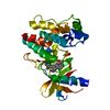 3dtcS C: citing same article ( S: Starting model for refinement |
|---|---|
| Similar structure data |

-Links
 PDBj
PDBj


- Assembly
| Deposited unit | 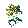
| ||||||||
|---|---|---|---|---|---|---|---|---|---|
| 1 |
| ||||||||
| Unit cell |
|
-Components
| #1: Protein | Mass: 32616.539 Da / Num. of mol.: 1 / Fragment: Roco4 Kinase Domain, UNP residues 1018-1292 Source method: isolated from a genetically manipulated source Source: (gene. exp.)  References: UniProt: Q6XHB2, non-specific serine/threonine protein kinase |
|---|---|
| #2: Water | ChemComp-HOH /  Mass: 18.015 Da / Num. of mol.: 62 / Source method: isolated from a natural source / Formula: H2O Mass: 18.015 Da / Num. of mol.: 62 / Source method: isolated from a natural source / Formula: H2O |
-Experimental details
-Experiment
| Experiment | Method: X-RAY DIFFRACTION / Number of used crystals: 1 |
|---|
- Sample preparation
| Crystal | Density Matthews: 2.45 Å3/Da / Density % sol: 49.93 % |
|---|---|
| Crystal grow | Temperature: 277 K / Method: vapor diffusion, hanging drop / pH: 8.5 Details: 0.1 M Bis- Tris, 0.2 M Na/K Tartrate; 11% PEG 3350, pH 8.5, vapor diffusion, hanging drop, temperature 277K |
-Data collection
| Diffraction | Mean temperature: 100 K | |||||||||||||||||||||||||||||||||||||||||||||||||||||||||||||||||||||||||||||||||||||||||||||||||||||||||||||||||||||||||||||||||||||||||||||||||||
|---|---|---|---|---|---|---|---|---|---|---|---|---|---|---|---|---|---|---|---|---|---|---|---|---|---|---|---|---|---|---|---|---|---|---|---|---|---|---|---|---|---|---|---|---|---|---|---|---|---|---|---|---|---|---|---|---|---|---|---|---|---|---|---|---|---|---|---|---|---|---|---|---|---|---|---|---|---|---|---|---|---|---|---|---|---|---|---|---|---|---|---|---|---|---|---|---|---|---|---|---|---|---|---|---|---|---|---|---|---|---|---|---|---|---|---|---|---|---|---|---|---|---|---|---|---|---|---|---|---|---|---|---|---|---|---|---|---|---|---|---|---|---|---|---|---|---|---|---|
| Diffraction source | Source: SYNCHROTRON / Site: SLS  / Beamline: X10SA / Wavelength: 0.9778 Å / Beamline: X10SA / Wavelength: 0.9778 Å | |||||||||||||||||||||||||||||||||||||||||||||||||||||||||||||||||||||||||||||||||||||||||||||||||||||||||||||||||||||||||||||||||||||||||||||||||||
| Detector | Type: DECTRIS PILATUS 6M / Detector: PIXEL / Date: Mar 20, 2011 | |||||||||||||||||||||||||||||||||||||||||||||||||||||||||||||||||||||||||||||||||||||||||||||||||||||||||||||||||||||||||||||||||||||||||||||||||||
| Radiation | Protocol: SINGLE WAVELENGTH / Monochromatic (M) / Laue (L): M / Scattering type: x-ray | |||||||||||||||||||||||||||||||||||||||||||||||||||||||||||||||||||||||||||||||||||||||||||||||||||||||||||||||||||||||||||||||||||||||||||||||||||
| Radiation wavelength | Wavelength: 0.9778 Å / Relative weight: 1 | |||||||||||||||||||||||||||||||||||||||||||||||||||||||||||||||||||||||||||||||||||||||||||||||||||||||||||||||||||||||||||||||||||||||||||||||||||
| Reflection | Resolution: 2→19.2 Å / Num. all: 23425 / Num. obs: 23425 / % possible obs: 100 % / Observed criterion σ(F): -3 / Observed criterion σ(I): -3 / Biso Wilson estimate: 36.487 Å2 / Rmerge(I) obs: 0.104 / Net I/σ(I): 13.48 | |||||||||||||||||||||||||||||||||||||||||||||||||||||||||||||||||||||||||||||||||||||||||||||||||||||||||||||||||||||||||||||||||||||||||||||||||||
| Reflection shell | Diffraction-ID: 1
|
- Processing
| Software |
| |||||||||||||||||||||||||||||||||||||||||||||
|---|---|---|---|---|---|---|---|---|---|---|---|---|---|---|---|---|---|---|---|---|---|---|---|---|---|---|---|---|---|---|---|---|---|---|---|---|---|---|---|---|---|---|---|---|---|---|
| Refinement | Method to determine structure: MOLECULAR REPLACEMENT Starting model: 3DTC Resolution: 2→19.2 Å / Cor.coef. Fo:Fc: 0.936 / Cor.coef. Fo:Fc free: 0.913 / Occupancy max: 1 / Occupancy min: 1 / SU B: 4.301 / SU ML: 0.122 / Cross valid method: THROUGHOUT / σ(F): 0 / ESU R: 0.191 / ESU R Free: 0.171 / Stereochemistry target values: MAXIMUM LIKELIHOOD Details: HYDROGENS HAVE BEEN USED IF PRESENT IN THE INPUT U VALUES : REFINED INDIVIDUALLY
| |||||||||||||||||||||||||||||||||||||||||||||
| Solvent computation | Ion probe radii: 0.8 Å / Shrinkage radii: 0.8 Å / VDW probe radii: 1.2 Å / Solvent model: MASK | |||||||||||||||||||||||||||||||||||||||||||||
| Displacement parameters | Biso max: 117.29 Å2 / Biso mean: 43.3138 Å2 / Biso min: 18.35 Å2
| |||||||||||||||||||||||||||||||||||||||||||||
| Refinement step | Cycle: LAST / Resolution: 2→19.2 Å
| |||||||||||||||||||||||||||||||||||||||||||||
| Refine LS restraints |
| |||||||||||||||||||||||||||||||||||||||||||||
| LS refinement shell | Resolution: 2→2.051 Å / Total num. of bins used: 20
|
