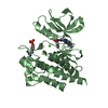
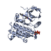
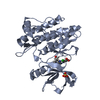
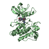
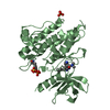
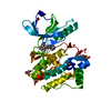
[English] 日本語
Yorodumi
- PDB-4xli: Crystal structure of Abl2/Arg kinase in complex with dasatinib -

+
Open data


ID or keywords:

Loading...

-
Basic information

Entry
Database: PDB / ID: 4xli
TitleCrystal structure of Abl2/Arg kinase in complex with dasatinib
ComponentsNon-specific protein-tyrosine kinaseNon-receptor tyrosine kinase
Keywordstransferase/transferase inhibitor / Transferase / inhibitor / kinase / transferase-transferase inhibitor complex
Function / homology
Function and homology information


circulatory system development => GO:0072359 / auditory behavior / Role of ABL in ROBO-SLIT signaling / reproductive process / aggressive behavior / protein localization => GO:0008104 / positive regulation of oxidoreductase activity / neuroepithelial cell differentiation / substrate-dependent cell migration, cell extension / positive regulation of Wnt signaling pathway, planar cell polarity pathway ...circulatory system development => GO:0072359 / auditory behavior / Role of ABL in ROBO-SLIT signaling / reproductive process / aggressive behavior / protein localization => GO:0008104 / positive regulation of oxidoreductase activity / neuroepithelial cell differentiation / substrate-dependent cell migration, cell extension / positive regulation of Wnt signaling pathway, planar cell polarity pathway / regulation of extracellular matrix organization / cerebellum morphogenesis / adult walking behavior / dendrite morphogenesis / alpha-beta T cell differentiation / exploration behavior / dendritic spine maintenance / negative regulation of Rho protein signal transduction / phagocytic cup / neuron remodeling / negative regulation of cell-cell adhesion / actin filament bundle assembly / Bergmann glial cell differentiation / neuromuscular process controlling balance / platelet-derived growth factor receptor signaling pathway / positive regulation of phospholipase C activity / phagocytosis / negative regulation of endothelial cell apoptotic process / cellular response to retinoic acid / positive regulation of interleukin-2 production / phosphotyrosine residue binding / post-embryonic development / learning / actin filament organization / neural tube closure / non-specific protein-tyrosine kinase / multicellular organism growth / non-membrane spanning protein tyrosine kinase activity / visual learning / epidermal growth factor receptor signaling pathway / positive regulation of neuron projection development / neuron differentiation / peptidyl-tyrosine phosphorylation / positive regulation of type II interferon production / actin cytoskeleton / lamellipodium / manganese ion binding / positive regulation of protein binding / positive regulation of cytosolic calcium ion concentration / cytoplasmic vesicle / actin cytoskeleton organization / protein tyrosine kinase activity / positive regulation of canonical NF-kappaB signal transduction / dendritic spine / positive regulation of ERK1 and ERK2 cascade / cell adhesion / protein phosphorylation / magnesium ion binding / ATP binding
Similarity search - Function
F-actin binding / F-actin binding / F-actin binding domain (FABD) / Tyrosine-protein kinase ABL, SH2 domain / SH3 domain / SH2 domain / Src homology 2 (SH2) domain profile. / Src homology 2 domains / SH2 domain / Src homology 3 domains ...F-actin binding / F-actin binding / F-actin binding domain (FABD) / Tyrosine-protein kinase ABL, SH2 domain / SH3 domain / SH2 domain / Src homology 2 (SH2) domain profile. / Src homology 2 domains / SH2 domain / Src homology 3 domains / SH2 domain superfamily / SH3-like domain superfamily / Src homology 3 (SH3) domain profile. / SH3 domain / Tyrosine-protein kinase, catalytic domain / Tyrosine kinase, catalytic domain / Tyrosine protein kinases specific active-site signature. / Tyrosine-protein kinase, active site / Protein tyrosine and serine/threonine kinase / Serine-threonine/tyrosine-protein kinase, catalytic domain / Transferase(Phosphotransferase) domain 1 / Transferase(Phosphotransferase); domain 1 / Phosphorylase Kinase; domain 1 / Phosphorylase Kinase; domain 1 / Protein kinase, ATP binding site / Protein kinases ATP-binding region signature. / Protein kinase domain profile. / Protein kinase domain / Protein kinase-like domain superfamily / 2-Layer Sandwich / Orthogonal Bundle / Mainly Alpha / Alpha Beta
Similarity search - Domain/homology
Chem-1N1 / Tyrosine-protein kinase / Tyrosine-protein kinase ABL2
Similarity search - Component
Biological speciesMus musculus (house mouse)
MethodX-RAY DIFFRACTION / SYNCHROTRON / MOLECULAR REPLACEMENT / Resolution: 2.5 Å
AuthorsHa, B.H. / Boggon, T.J.
Funding support United States, 1items
OrganizationGrant numberCountry
National Institutes of Health/National Institute of General Medical Sciences (NIH/NIGMS)R01GM100411 United States
CitationJournal: Acta Crystallogr.,Sect.F / Year: 2015
Title: Structure of the ABL2/ARG kinase in complex with dasatinib.
Authors: Ha, B.H. / Simpson, M.A. / Koleske, A.J. / Boggon, T.J.
History
DepositionJan 13, 2015Deposition site: RCSB / Processing site: RCSB
Revision 1.0Apr 1, 2015Provider: repository / Type: Initial release
Revision 1.1Jun 24, 2015Group: Database references
Revision 1.2Sep 13, 2017Group: Author supporting evidence / Derived calculations / Source and taxonomy
Category: entity_src_gen / pdbx_audit_support / pdbx_struct_oper_list
Item: _entity_src_gen.pdbx_alt_source_flag / _pdbx_audit_support.funding_organization / _pdbx_struct_oper_list.symmetry_operation
Revision 1.3Dec 25, 2019Group: Author supporting evidence / Category: pdbx_audit_support / Item: _pdbx_audit_support.funding_organization
Revision 1.4Sep 27, 2023Group: Data collection / Database references ...Data collection / Database references / Derived calculations / Refinement description
Category: chem_comp_atom / chem_comp_bond ...chem_comp_atom / chem_comp_bond / database_2 / pdbx_initial_refinement_model / struct_conn
Item: _database_2.pdbx_DOI / _database_2.pdbx_database_accession ..._database_2.pdbx_DOI / _database_2.pdbx_database_accession / _struct_conn.ptnr1_auth_asym_id / _struct_conn.ptnr1_auth_comp_id / _struct_conn.ptnr1_auth_seq_id / _struct_conn.ptnr1_label_asym_id / _struct_conn.ptnr1_label_atom_id / _struct_conn.ptnr1_label_comp_id / _struct_conn.ptnr1_label_seq_id / _struct_conn.ptnr2_auth_asym_id / _struct_conn.ptnr2_auth_comp_id / _struct_conn.ptnr2_auth_seq_id / _struct_conn.ptnr2_label_asym_id / _struct_conn.ptnr2_label_atom_id / _struct_conn.ptnr2_label_comp_id / _struct_conn.ptnr2_label_seq_id

-
Structure visualization

Structure viewerMolecule:
MolmilJmol/JSmol

Downloads & links

-
Assembly

Deposited unit
A: Non-specific protein-tyrosine kinase
B: Non-specific protein-tyrosine kinase
hetero molecules


Theoretical massNumber of molelcules
Total (without water)62,5805
Polymers61,5392
Non-polymers1,0413
Water57632
1
A: Non-specific protein-tyrosine kinase
hetero molecules


Theoretical massNumber of molelcules
Total (without water)31,3233
Polymers30,7691
Non-polymers5532
Water181
TypeNameSymmetry operationNumber
identity operation1_555x,y,z1
2
B: Non-specific protein-tyrosine kinase
hetero molecules


Theoretical massNumber of molelcules
Total (without water)31,2572
Polymers30,7691
Non-polymers4881
Water181
TypeNameSymmetry operationNumber
identity operation1_555x,y,z1
Unit cell
Length a, b, c (Å)109.688, 109.688, 121.520
Angle α, β, γ (deg.)90.00, 90.00, 120.00
Int Tables number170
Space group name H-MP65

-
Components

#1: Protein Non-specific protein-tyrosine kinase / Non-receptor tyrosine kinase


Mass: 30769.336 Da / Num. of mol.: 2
Source method: isolated from a genetically manipulated source
Source: (gene. exp.) Mus musculus (house mouse) / Gene: Abl2 / Plasmid: pFastBac-HTA / Production host: Trichoplusia ni (cabbage looper) / Strain (production host): Hi 5
References: UniProt: F8VQH0, UniProt: Q4JIM5*PLUS, non-specific protein-tyrosine kinase
#2: Chemical ChemComp-1N1 / N-(2-CHLORO-6-METHYLPHENYL)-2-({6-[4-(2-HYDROXYETHYL)PIPERAZIN-1-YL]-2-METHYLPYRIMIDIN-4-YL}AMINO)-1,3-THIAZOLE-5-CARBOXAMIDE / Dasatinib / Dasatinib


Mass: 488.006 Da / Num. of mol.: 2 / Source method: obtained synthetically / Formula: C22H26ClN7O2S / Comment: medication*YM
#3: Chemical ChemComp-ZN / ZINC ION


Mass: 65.409 Da / Num. of mol.: 1 / Source method: obtained synthetically / Formula: Zn
#4: Water ChemComp-HOH / water / Water


Mass: 18.015 Da / Num. of mol.: 32 / Source method: isolated from a natural source / Formula: H2O

-
Experimental details

-
Experiment

ExperimentMethod: X-RAY DIFFRACTION / Number of used crystals: 1

-
Sample preparation

CrystalDensity Matthews: 3.38 Å3/Da / Density % sol: 63.6 %
Crystal growTemperature: 295 K / Method: vapor diffusion, hanging drop / pH: 7
Details: 0.1 M HEPES pH 7.0, 1.3M Sodium Malonate, 2.5 % Jeffamine ED-2001
PH range: 6.5-7.5 / Temp details: Room temperature

-
Data collection

DiffractionMean temperature: 83 K / Ambient temp details: Cryo
Diffraction sourceSource: SYNCHROTRON / Site: APS / Beamline: 24-ID-E / Wavelength: 0.97916 Å
DetectorType: ADSC QUANTUM 315 / Detector: CCD / Date: Jun 7, 2014
RadiationMonochromator: Si(111) / Protocol: SINGLE WAVELENGTH / Monochromatic (M) / Laue (L): M / Scattering type: x-ray
Radiation wavelengthWavelength: 0.97916 Å / Relative weight: 1
Reflection twin
Crystal-IDIDOperatorDomain-IDFraction
11H, K, L10.712
11K, H, -L20.288
ReflectionResolution: 2.5→47.5 Å / Num. obs: 29610 / % possible obs: 100 % / Observed criterion σ(F): 0 / Observed criterion σ(I): -3 / Redundancy: 11.1 % / Rsym value: 0.241 / Net I/σ(I): 8.5
Reflection shellResolution: 2.5→2.59 Å / Redundancy: 11 % / Mean I/σ(I) obs: 1.17 / % possible all: 100

-
Processing

Software
NameVersionClassification
REFMAC5.8.0071refinement
PHASERphasing
HKL-2000data reduction
Cootmodel building
HKL-2000data scaling
RefinementMethod to determine structure: MOLECULAR REPLACEMENT
Starting model: 2XYN
Resolution: 2.5→47.5 Å / Cor.coef. Fo:Fc: 0.964 / Cor.coef. Fo:Fc free: 0.939 / SU B: 8.866 / SU ML: 0.102 / Cross valid method: THROUGHOUT / ESU R: 0.055 / ESU R Free: 0.044 / Stereochemistry target values: MAXIMUM LIKELIHOOD / Details: HYDROGENS HAVE BEEN ADDED IN THE RIDING POSITIONS
RfactorNum. reflection% reflectionSelection details
Rfree0.205 1478 5.1 %RANDOM
Rwork0.154 ---
obs0.157 27244 99.98 %-
Solvent computationIon probe radii: 0.8 Å / Shrinkage radii: 0.8 Å / VDW probe radii: 1.2 Å / Solvent model: MASK
Displacement parametersBiso mean: 48.8 Å2
Baniso -1Baniso -2Baniso -3
1-14.99 Å20 Å20 Å2
2--14.99 Å20 Å2
3----29.99 Å2
Refinement stepCycle: 1 / Resolution: 2.5→47.5 Å
ProteinNucleic acidLigandSolventTotal
Num. atoms4259 0 67 32 4358
Refine LS restraints
Refine-IDTypeDev idealDev ideal targetNumber
X-RAY DIFFRACTIONr_bond_refined_d0.0190.0194446
X-RAY DIFFRACTIONr_bond_other_d0.0020.024155
X-RAY DIFFRACTIONr_angle_refined_deg2.0151.9756031
X-RAY DIFFRACTIONr_angle_other_deg1.05739600
X-RAY DIFFRACTIONr_dihedral_angle_1_deg7.2865524
X-RAY DIFFRACTIONr_dihedral_angle_2_deg36.64524.01192
X-RAY DIFFRACTIONr_dihedral_angle_3_deg15.71815756
X-RAY DIFFRACTIONr_dihedral_angle_4_deg12.8421518
X-RAY DIFFRACTIONr_chiral_restr0.1110.2645
X-RAY DIFFRACTIONr_gen_planes_refined0.010.0214892
X-RAY DIFFRACTIONr_gen_planes_other0.0080.021002
X-RAY DIFFRACTIONr_nbd_refined
X-RAY DIFFRACTIONr_nbd_other
X-RAY DIFFRACTIONr_nbtor_refined
X-RAY DIFFRACTIONr_nbtor_other
X-RAY DIFFRACTIONr_xyhbond_nbd_refined
X-RAY DIFFRACTIONr_xyhbond_nbd_other
X-RAY DIFFRACTIONr_metal_ion_refined
X-RAY DIFFRACTIONr_metal_ion_other
X-RAY DIFFRACTIONr_symmetry_vdw_refined
X-RAY DIFFRACTIONr_symmetry_vdw_other
X-RAY DIFFRACTIONr_symmetry_hbond_refined
X-RAY DIFFRACTIONr_symmetry_hbond_other
X-RAY DIFFRACTIONr_symmetry_metal_ion_refined
X-RAY DIFFRACTIONr_symmetry_metal_ion_other
X-RAY DIFFRACTIONr_mcbond_it4.2364.3422110
X-RAY DIFFRACTIONr_mcbond_other4.2364.3422108
X-RAY DIFFRACTIONr_mcangle_it6.0416.5042628
X-RAY DIFFRACTIONr_mcangle_other6.046.5042629
X-RAY DIFFRACTIONr_scbond_it4.9324.7642336
X-RAY DIFFRACTIONr_scbond_other4.9314.7642336
X-RAY DIFFRACTIONr_scangle_it
X-RAY DIFFRACTIONr_scangle_other7.0716.9763404
X-RAY DIFFRACTIONr_long_range_B_refined8.91335.0285073
X-RAY DIFFRACTIONr_long_range_B_other8.91235.0385074
X-RAY DIFFRACTIONr_rigid_bond_restr
X-RAY DIFFRACTIONr_sphericity_free
X-RAY DIFFRACTIONr_sphericity_bonded
LS refinement shellResolution: 2.5→2.565 Å / Total num. of bins used: 20
RfactorNum. reflection% reflection
Rfree0.376 103 -
Rwork0.262 2012 -
obs--99.86 %
Refinement TLS params.

Method: refined / Refine-ID: X-RAY DIFFRACTION

IDL112)L122)L132)L222)L232)L332)S11 (Å °)S12 (Å °)S13 (Å °)S21 (Å °)S22 (Å °)S23 (Å °)S31 (Å °)S32 (Å °)S33 (Å °)T112)T122)T132)T222)T232)T332)Origin x (Å)Origin y (Å)Origin z (Å)
11.5516-0.50420.03481.44670.26760.7028-0.1086-0.2065-0.08160.13090.00130.30560.2162-0.14610.10730.0776-0.0280.03480.0576-0.01920.092641.50819.3702-7.0358
21.6386-0.24850.18030.98720.34860.8193-0.08230.06320.3238-0.1582-0.05120.1058-0.17990.10430.13350.0539-0.0176-0.04140.02960.00010.117960.601748.1829-18.9481
Refinement TLS group
IDRefine-IDRefine TLS-IDAuth asym-IDAuth seq-ID
1X-RAY DIFFRACTION1A279 - 291
2X-RAY DIFFRACTION1A292 - 308
3X-RAY DIFFRACTION1A309 - 348
4X-RAY DIFFRACTION1A349 - 360
5X-RAY DIFFRACTION1A361 - 546
6X-RAY DIFFRACTION2B279 - 291
7X-RAY DIFFRACTION2B292 - 308
8X-RAY DIFFRACTION2B309 - 348
9X-RAY DIFFRACTION2B349 - 360
10X-RAY DIFFRACTION2B361 - 546

+
About Yorodumi

-
News

-
Feb 9, 2022. New format data for meta-information of EMDB entries

New format data for meta-information of EMDB entries

  • Version 3 of the EMDB header file is now the official format.
  • The previous official version 1.9 will be removed from the archive.

Related info.:EMDB header

External links:wwPDB to switch to version 3 of the EMDB data model

-
Aug 12, 2020. Covid-19 info

Covid-19 info

URL: https://pdbj.org/emnavi/covid19.php

New page: Covid-19 featured information page in EM Navigator.

Related info.:Covid-19 info / Mar 5, 2020. Novel coronavirus structure data

+
Mar 5, 2020. Novel coronavirus structure data

Novel coronavirus structure data

Related info.:Yorodumi Speices / Aug 12, 2020. Covid-19 info

External links:COVID-19 featured content - PDBj / Molecule of the Month (242):Coronavirus Proteases

+
Jan 31, 2019. EMDB accession codes are about to change! (news from PDBe EMDB page)

EMDB accession codes are about to change! (news from PDBe EMDB page)

  • The allocation of 4 digits for EMDB accession codes will soon come to an end. Whilst these codes will remain in use, new EMDB accession codes will include an additional digit and will expand incrementally as the available range of codes is exhausted. The current 4-digit format prefixed with “EMD-” (i.e. EMD-XXXX) will advance to a 5-digit format (i.e. EMD-XXXXX), and so on. It is currently estimated that the 4-digit codes will be depleted around Spring 2019, at which point the 5-digit format will come into force.
  • The EM Navigator/Yorodumi systems omit the EMD- prefix.

Related info.:Q: What is EMD? / ID/Accession-code notation in Yorodumi/EM Navigator

External links:EMDB Accession Codes are Changing Soon! / Contact to PDBj

+
Jul 12, 2017. Major update of PDB

Major update of PDB

  • wwPDB released updated PDB data conforming to the new PDBx/mmCIF dictionary.
  • This is a major update changing the version number from 4 to 5, and with Remediation, in which all the entries are updated.
  • In this update, many items about electron microscopy experimental information are reorganized (e.g. em_software).
  • Now, EM Navigator and Yorodumi are based on the updated data.

External links:wwPDB Remediation / Enriched Model Files Conforming to OneDep Data Standards Now Available in the PDB FTP Archive

-
Yorodumi

Thousand views of thousand structures

  • Yorodumi is a browser for structure data from EMDB, PDB, SASBDB, etc.
  • This page is also the successor to EM Navigator detail page, and also detail information page/front-end page for Omokage search.
  • The word "yorodu" (or yorozu) is an old Japanese word meaning "ten thousand". "mi" (miru) is to see.

Related info.:EMDB / PDB / SASBDB / Comparison of 3 databanks / Yorodumi Search / Aug 31, 2016. New EM Navigator & Yorodumi / Yorodumi Papers / Jmol/JSmol / Function and homology information / Changes in new EM Navigator and Yorodumi

Read more