 Movie
Movie Controller

[English] 日本語
 Yorodumi
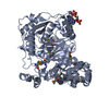
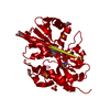
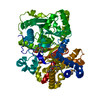
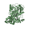
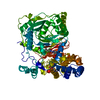
- PDB-4ep9: CRYSTAL STRUCTURE OF RAT CARNITINE PALMITOYLTRANSFERASE 2 IN COMP... -
+ Open data

- Basic information
| Entry | Database: PDB / ID: 4ep9 | ||||||
|---|---|---|---|---|---|---|---|
| Title | CRYSTAL STRUCTURE OF RAT CARNITINE PALMITOYLTRANSFERASE 2 IN COMPLEX WITH CoA-site inhibitor | ||||||
 Components Components | Carnitine O-palmitoyltransferase 2, mitochondrial | ||||||
 Keywords Keywords | TRANSFERASE/TRANSFERASE INHIBITOR / TRANSFERASE / ACYLTRANSFERASE / MITOCHONDRIAL PROTEIN / CoA / acylcarnitine / mitochondrial inner membrane / LIPID TRANSPORT / TRANSFERASE-TRANSFERASE INHIBITOR complex | ||||||
| Function / homology |  Function and homology information Function and homology informationcarnitine O-palmitoyltransferase / carnitine O-palmitoyltransferase activity / carnitine shuttle / Carnitine shuttle / carnitine O-octanoyltransferase activity / carnitine metabolic process / long-chain fatty acid metabolic process / response to fatty acid / acyltransferase activity / fatty acid beta-oxidation ...carnitine O-palmitoyltransferase / carnitine O-palmitoyltransferase activity / carnitine shuttle / Carnitine shuttle / carnitine O-octanoyltransferase activity / carnitine metabolic process / long-chain fatty acid metabolic process / response to fatty acid / acyltransferase activity / fatty acid beta-oxidation / long-chain fatty acid transport / positive regulation of cold-induced thermogenesis / in utero embryonic development / mitochondrial inner membrane / mitochondrial matrix / nucleolus / mitochondrion / nucleoplasm Similarity search - Function | ||||||
| Biological species |  | ||||||
| Method |  X-RAY DIFFRACTION / SYNCHROTRON / MOLECULAR REPLACEMENT / Resolution: 2.03 Å X-RAY DIFFRACTION / SYNCHROTRON / MOLECULAR REPLACEMENT / Resolution: 2.03 Å | ||||||
 Authors Authors | Rufer, A.C. / Thoma, R. / Benz, J. / Stihle, M. / Gsell, B. / De Roo, E. / Banner, D.W. / Mueller, F. / Chomienne, O. / Hennig, M. | ||||||
 Citation Citation | Journal: FEBS Open Bio / Year: 2013 Title: Isothermal titration calorimetry with micelles: Thermodynamics of inhibitor binding to carnitine palmitoyltransferase 2 membrane protein. Authors: Perspicace, S. / Rufer, A.C. / Thoma, R. / Mueller, F. / Hennig, M. / Ceccarelli, S. / Schulz-Gasch, T. / Seelig, J. | ||||||
| History |
|
- Structure visualization
| Structure viewer | Molecule: MolmilJmol/JSmol |
|---|
- Downloads & links
-Download
| PDBx/mmCIF format | 4ep9.cif.gz | 150.3 KB | Display | PDBx/mmCIF format |
|---|---|---|---|---|
| PDB format | pdb4ep9.ent.gz | 114.5 KB | Display | PDB format |
| PDBx/mmJSON format | 4ep9.json.gz | Tree view | PDBx/mmJSON format | |
| Others |  Other downloads Other downloads |
-Validation report
| Arichive directory | https://data.pdbj.org/pub/pdb/validation_reports/ep/4ep9ftp://data.pdbj.org/pub/pdb/validation_reports/ep/4ep9 | HTTPS FTP |
|---|
-Related structure data
| Related structure data |  4ephC  4eywC  2debS S: Starting model for refinement C: citing same article ( |
|---|---|
| Similar structure data |


-Links
 PDBj
PDBj


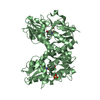
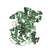
- Assembly
| Deposited unit | 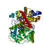
| ||||||||
|---|---|---|---|---|---|---|---|---|---|
| 1 |
| ||||||||
| Unit cell |
|
-Components
| #1: Protein | Mass: 73566.094 Da / Num. of mol.: 1 / Fragment: Carnitine O-palmitoyltransferase 2, mitochondrial Source method: isolated from a genetically manipulated source Details: pET28a_rCPT-2_27-658 / Source: (gene. exp.)  References: UniProt: P18886, carnitine O-palmitoyltransferase |
|---|---|
| #2: Chemical | ChemComp-0RD /   Mass: 500.951 Da / Num. of mol.: 1 / Source method: obtained synthetically / Formula: C24H21ClN2O6S Mass: 500.951 Da / Num. of mol.: 1 / Source method: obtained synthetically / Formula: C24H21ClN2O6S |
| #3: Chemical | ChemComp-PLM /   Mass: 256.424 Da / Num. of mol.: 1 / Source method: obtained synthetically / Formula: C16H32O2 Mass: 256.424 Da / Num. of mol.: 1 / Source method: obtained synthetically / Formula: C16H32O2 |
| #4: Water | ChemComp-HOH /  Mass: 18.015 Da / Num. of mol.: 407 / Source method: isolated from a natural source / Formula: H2O Mass: 18.015 Da / Num. of mol.: 407 / Source method: isolated from a natural source / Formula: H2O |
-Experimental details
-Experiment
| Experiment | Method: X-RAY DIFFRACTION / Number of used crystals: 1 |
|---|
- Sample preparation
| Crystal | Density Matthews: 2.39 Å3/Da / Density % sol: 48.57 % |
|---|---|
| Crystal grow | Temperature: 293 K / Method: vapor diffusion, hanging drop / pH: 7.5 Details: Hampton Index #76 0.2 M lithium sulfate monohydrate, 0.1 M HEPES pH 7.5, 25 % (w/v) polyethylene glycol 3,350 , VAPOR DIFFUSION, HANGING DROP, temperature 293K |
-Data collection
| Diffraction | Mean temperature: 80 K |
|---|---|
| Diffraction source | Source: SYNCHROTRON / Site: SLS  / Beamline: X10SA / Wavelength: 0.97955 Å / Beamline: X10SA / Wavelength: 0.97955 Å |
| Detector | Type: MARRESEARCH / Detector: CCD / Date: Dec 13, 2005 |
| Radiation | Monochromator: LN2 COOLED FIXED-EXIT SI(111) MONOCHROMATOR (19.65M, FOCUSING SAGITTAL-HORIZONTAL) BENDABLE MIRROR (20.50 M FOCUSING MERIDIONAL-VERTICAL) Protocol: SINGLE WAVELENGTH / Monochromatic (M) / Laue (L): M / Scattering type: x-ray |
| Radiation wavelength | Wavelength: 0.97955 Å / Relative weight: 1 |
| Reflection | Resolution: 2.03→19.7 Å / Num. all: 47020 / % possible obs: 98.4 % / Observed criterion σ(I): 3.42 |
| Reflection shell | Resolution: 2.03→2.15 Å / % possible all: 91.4 |
- Processing
| Software |
| ||||||||||||||||||||||||||||||||||||||||||||||||||||||||||||||||||||||||||||||||||||||||||
|---|---|---|---|---|---|---|---|---|---|---|---|---|---|---|---|---|---|---|---|---|---|---|---|---|---|---|---|---|---|---|---|---|---|---|---|---|---|---|---|---|---|---|---|---|---|---|---|---|---|---|---|---|---|---|---|---|---|---|---|---|---|---|---|---|---|---|---|---|---|---|---|---|---|---|---|---|---|---|---|---|---|---|---|---|---|---|---|---|---|---|---|
| Refinement | Method to determine structure: MOLECULAR REPLACEMENT Starting model: PDB ENTRY 2DEB Resolution: 2.03→19.7 Å / Cor.coef. Fo:Fc: 0.944 / Cor.coef. Fo:Fc free: 0.898 / SU B: 4.311 / SU ML: 0.12 / Cross valid method: THROUGHOUT / ESU R: 0.18 / ESU R Free: 0.175 / Stereochemistry target values: MAXIMUM LIKELIHOOD / Details: HYDROGENS HAVE BEEN ADDED IN THE RIDING POSITIONS
| ||||||||||||||||||||||||||||||||||||||||||||||||||||||||||||||||||||||||||||||||||||||||||
| Solvent computation | Ion probe radii: 0.8 Å / Shrinkage radii: 0.8 Å / VDW probe radii: 1.2 Å / Solvent model: MASK | ||||||||||||||||||||||||||||||||||||||||||||||||||||||||||||||||||||||||||||||||||||||||||
| Displacement parameters | Biso mean: 21.734 Å2
| ||||||||||||||||||||||||||||||||||||||||||||||||||||||||||||||||||||||||||||||||||||||||||
| Refinement step | Cycle: LAST / Resolution: 2.03→19.7 Å
| ||||||||||||||||||||||||||||||||||||||||||||||||||||||||||||||||||||||||||||||||||||||||||
| Refine LS restraints |
| ||||||||||||||||||||||||||||||||||||||||||||||||||||||||||||||||||||||||||||||||||||||||||
| LS refinement shell | Resolution: 2.03→2.08 Å / Total num. of bins used: 20
|
