 Movie
Movie Controller

[English] 日本語
 Yorodumi
- PDB-3pmv: Ligand-binding domain of GluA2 (flip) ionotropic glutamate recept... -
+ Open data

- Basic information
| Entry | Database: PDB / ID: 3pmv | ||||||
|---|---|---|---|---|---|---|---|
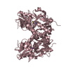
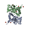
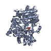
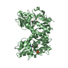
| Title | Ligand-binding domain of GluA2 (flip) ionotropic glutamate receptor in complex with an allosteric modulator | ||||||
 Components Components | Glutamate receptor 2 | ||||||
 Keywords Keywords | TRANSPORT PROTEIN / Membrane protein / Fusion protein / chimera protein | ||||||
| Function / homology |  Function and homology information Function and homology informationspine synapse / dendritic spine neck / dendritic spine cytoplasm / dendritic spine head / cellular response to amine stimulus / Activation of AMPA receptors / ligand-gated monoatomic cation channel activity / perisynaptic space / Trafficking of GluR2-containing AMPA receptors / response to lithium ion ...spine synapse / dendritic spine neck / dendritic spine cytoplasm / dendritic spine head / cellular response to amine stimulus / Activation of AMPA receptors / ligand-gated monoatomic cation channel activity / perisynaptic space / Trafficking of GluR2-containing AMPA receptors / response to lithium ion / AMPA glutamate receptor activity / AMPA glutamate receptor clustering / kainate selective glutamate receptor activity / immunoglobulin binding / AMPA glutamate receptor complex / regulation of receptor recycling / extracellularly glutamate-gated ion channel activity / cellular response to glycine / ionotropic glutamate receptor complex / asymmetric synapse / Unblocking of NMDA receptors, glutamate binding and activation / glutamate receptor binding / positive regulation of synaptic transmission / conditioned place preference / response to fungicide / regulation of synaptic transmission, glutamatergic / extracellular ligand-gated monoatomic ion channel activity / cytoskeletal protein binding / glutamate-gated receptor activity / cellular response to brain-derived neurotrophic factor stimulus / regulation of long-term synaptic depression / somatodendritic compartment / glutamate-gated calcium ion channel activity / presynaptic active zone membrane / excitatory synapse / ionotropic glutamate receptor signaling pathway / ionotropic glutamate receptor binding / dendrite cytoplasm / dendrite membrane / ligand-gated monoatomic ion channel activity involved in regulation of presynaptic membrane potential / positive regulation of excitatory postsynaptic potential / dendritic shaft / SNARE binding / synaptic membrane / PDZ domain binding / establishment of protein localization / protein tetramerization / synaptic transmission, glutamatergic / transmitter-gated monoatomic ion channel activity involved in regulation of postsynaptic membrane potential / cerebral cortex development / receptor internalization / postsynaptic density membrane / modulation of chemical synaptic transmission / Schaffer collateral - CA1 synapse / terminal bouton / synaptic vesicle / long-term synaptic potentiation / amyloid-beta binding / synaptic vesicle membrane / growth cone / presynapse / signaling receptor activity / presynaptic membrane / scaffold protein binding / dendritic spine / chemical synaptic transmission / perikaryon / postsynaptic membrane / neuron projection / postsynaptic density / external side of plasma membrane / axon / neuronal cell body / synapse / dendrite / protein kinase binding / protein-containing complex binding / glutamatergic synapse / cell surface / endoplasmic reticulum / protein-containing complex / membrane / identical protein binding / plasma membrane Similarity search - Function | ||||||
| Biological species |  | ||||||
| Method |  X-RAY DIFFRACTION / MOLECULAR REPLACEMENT / Resolution: 1.8 Å X-RAY DIFFRACTION / MOLECULAR REPLACEMENT / Resolution: 1.8 Å | ||||||
 Authors Authors | Maclean, J.K.F. / Jamieson, C. / Brown, C.I. / Campbell, R.A. / Gillen, K.J. / Gillespie, J. / Kazemier, B. / Kiczun, M. / Lamont, Y. / Lyons, A.J. ...Maclean, J.K.F. / Jamieson, C. / Brown, C.I. / Campbell, R.A. / Gillen, K.J. / Gillespie, J. / Kazemier, B. / Kiczun, M. / Lamont, Y. / Lyons, A.J. / Moir, E.M. / Morrow, J.A. / Pantling, J. / Rankovic, Z. / Smith, L. | ||||||
 Citation Citation | Journal: Bioorg.Med.Chem.Lett. / Year: 2011 Title: Structure based evolution of a novel series of positive modulators of the AMPA receptor. Authors: Jamieson, C. / Maclean, J.K. / Brown, C.I. / Campbell, R.A. / Gillen, K.J. / Gillespie, J. / Kazemier, B. / Kiczun, M. / Lamont, Y. / Lyons, A.J. / Moir, E.M. / Morrow, J.A. / Pantling, J. / ...Authors: Jamieson, C. / Maclean, J.K. / Brown, C.I. / Campbell, R.A. / Gillen, K.J. / Gillespie, J. / Kazemier, B. / Kiczun, M. / Lamont, Y. / Lyons, A.J. / Moir, E.M. / Morrow, J.A. / Pantling, J. / Rankovic, Z. / Smith, L. | ||||||
| History |
|
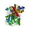
- Structure visualization
| Structure viewer | Molecule: MolmilJmol/JSmol |
|---|
- Downloads & links
-Download
| PDBx/mmCIF format | 3pmv.cif.gz | 79.6 KB | Display | PDBx/mmCIF format |
|---|---|---|---|---|
| PDB format | pdb3pmv.ent.gz | 58.9 KB | Display | PDB format |
| PDBx/mmJSON format | 3pmv.json.gz | Tree view | PDBx/mmJSON format | |
| Others |  Other downloads Other downloads |
-Validation report
| Arichive directory | https://data.pdbj.org/pub/pdb/validation_reports/pm/3pmvftp://data.pdbj.org/pub/pdb/validation_reports/pm/3pmv | HTTPS FTP |
|---|
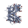
-Related structure data
| Related structure data | 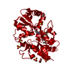 3pmwC 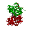 3pmxC 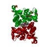 3o28S S: Starting model for refinement C: citing same article ( |
|---|---|
| Similar structure data |


-Links
 PDBj
PDBj


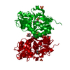
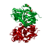
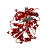
- Assembly
| Deposited unit | 
| ||||||||
|---|---|---|---|---|---|---|---|---|---|
| 1 | 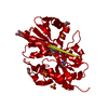
| ||||||||
| Unit cell |
|
-Components
-Protein , 1 types, 1 molecules A
| #1: Protein | Mass: 29177.670 Da / Num. of mol.: 1 Fragment: Ligand binding domain, residues 413 to 527 and 653 to 796 Source method: isolated from a genetically manipulated source Details: S1-S2 fusion in which Gly118 and Thr119 replace a membrane-spanning region Source: (gene. exp.)  |
|---|
-Non-polymers , 5 types, 370 molecules 

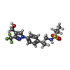


| #2: Chemical | ChemComp-GLU /  Type: L-peptide linking / Mass: 147.129 Da / Num. of mol.: 1 / Source method: obtained synthetically / Formula: C5H9NO4 Type: L-peptide linking / Mass: 147.129 Da / Num. of mol.: 1 / Source method: obtained synthetically / Formula: C5H9NO4 | ||||||
|---|---|---|---|---|---|---|---|
| #3: Chemical | ChemComp-SO4 /  Mass: 96.063 Da / Num. of mol.: 5 / Source method: obtained synthetically / Formula: SO4 Mass: 96.063 Da / Num. of mol.: 5 / Source method: obtained synthetically / Formula: SO4#4: Chemical | ChemComp-557 / |  Mass: 405.435 Da / Num. of mol.: 1 / Source method: obtained synthetically / Formula: C17H22F3N3O3S Mass: 405.435 Da / Num. of mol.: 1 / Source method: obtained synthetically / Formula: C17H22F3N3O3S#5: Chemical | ChemComp-GOL /  Mass: 92.094 Da / Num. of mol.: 4 / Source method: obtained synthetically / Formula: C3H8O3 Mass: 92.094 Da / Num. of mol.: 4 / Source method: obtained synthetically / Formula: C3H8O3#6: Water | ChemComp-HOH / | Mass: 18.015 Da / Num. of mol.: 359 / Source method: isolated from a natural source / Formula: H2O |
-Details
| Has protein modification | Y |
|---|
-Experimental details
-Experiment
| Experiment | Method: X-RAY DIFFRACTION / Number of used crystals: 1 |
|---|
- Sample preparation
| Crystal | Density Matthews: 2.27 Å3/Da / Density % sol: 45.91 % |
|---|---|
| Crystal grow | Temperature: 277 K / Method: vapor diffusion, hanging drop / pH: 4 Details: 18% PEG 4000, 50mM Lithium Sulphate, 2.5% Glycerol, 100mM Sodium Cacodylate pH 4.0, VAPOR DIFFUSION, HANGING DROP, temperature 277K |
-Data collection
| Diffraction | Mean temperature: 100 K |
|---|---|
| Diffraction source | Source: ROTATING ANODE / Type: RIGAKU MICROMAX-007 / Wavelength: 1.54178 Å |
| Detector | Type: RIGAKU RAXIS IV++ / Detector: IMAGE PLATE / Date: Mar 14, 2007 / Details: Mirrors |
| Radiation | Monochromator: Graphite / Protocol: SINGLE WAVELENGTH / Monochromatic (M) / Laue (L): M / Scattering type: x-ray |
| Radiation wavelength | Wavelength: 1.54178 Å / Relative weight: 1 |
| Reflection | Resolution: 1.79→43.61 Å / Num. all: 24891 / Num. obs: 24891 / % possible obs: 98.1 % / Observed criterion σ(I): 2 / Redundancy: 4.1 % / Biso Wilson estimate: 26.1 Å2 / Rmerge(I) obs: 0.05 / Net I/σ(I): 13.2 |
| Reflection shell | Resolution: 1.8→1.86 Å / Redundancy: 2.9 % / Rmerge(I) obs: 0.287 / Mean I/σ(I) obs: 3.5 / Num. unique all: 2050 / % possible all: 82.9 |
- Processing
| Software |
| |||||||||||||||||||||||||||||||||||||||||||||||||||||||||||||||||||||||||||||||||||||||||||||||||||||||||||||||||||||||||||||
|---|---|---|---|---|---|---|---|---|---|---|---|---|---|---|---|---|---|---|---|---|---|---|---|---|---|---|---|---|---|---|---|---|---|---|---|---|---|---|---|---|---|---|---|---|---|---|---|---|---|---|---|---|---|---|---|---|---|---|---|---|---|---|---|---|---|---|---|---|---|---|---|---|---|---|---|---|---|---|---|---|---|---|---|---|---|---|---|---|---|---|---|---|---|---|---|---|---|---|---|---|---|---|---|---|---|---|---|---|---|---|---|---|---|---|---|---|---|---|---|---|---|---|---|---|---|---|
| Refinement | Method to determine structure: MOLECULAR REPLACEMENT Starting model: 3O28 Resolution: 1.8→43.61 Å / Cor.coef. Fo:Fc: 0.96 / Cor.coef. Fo:Fc free: 0.928 / SU B: 3.149 / SU ML: 0.099 / Isotropic thermal model: Isotropic / Cross valid method: THROUGHOUT / ESU R Free: 0.155 / Stereochemistry target values: MAXIMUM LIKELIHOOD / Details: HYDROGENS HAVE BEEN ADDED IN THE RIDING POSITIONS
| |||||||||||||||||||||||||||||||||||||||||||||||||||||||||||||||||||||||||||||||||||||||||||||||||||||||||||||||||||||||||||||
| Solvent computation | Ion probe radii: 0.8 Å / Shrinkage radii: 0.8 Å / VDW probe radii: 1.4 Å / Solvent model: MASK | |||||||||||||||||||||||||||||||||||||||||||||||||||||||||||||||||||||||||||||||||||||||||||||||||||||||||||||||||||||||||||||
| Displacement parameters | Biso mean: 18.709 Å2
| |||||||||||||||||||||||||||||||||||||||||||||||||||||||||||||||||||||||||||||||||||||||||||||||||||||||||||||||||||||||||||||
| Refinement step | Cycle: LAST / Resolution: 1.8→43.61 Å
| |||||||||||||||||||||||||||||||||||||||||||||||||||||||||||||||||||||||||||||||||||||||||||||||||||||||||||||||||||||||||||||
| Refine LS restraints |
| |||||||||||||||||||||||||||||||||||||||||||||||||||||||||||||||||||||||||||||||||||||||||||||||||||||||||||||||||||||||||||||
| LS refinement shell | Resolution: 1.8→1.847 Å / Total num. of bins used: 20
|
