 Movie
Movie Controller

[English] 日本語
 Yorodumi
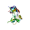
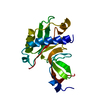
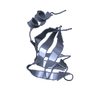
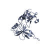
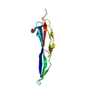
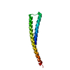
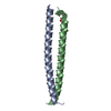
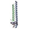
- PDB-4eo0: crystal structure of the pilus binding domain of the filamentous ... -
+ Open data

- Basic information
| Entry | Database: PDB / ID: 4eo0 | ||||||
|---|---|---|---|---|---|---|---|
| Title | crystal structure of the pilus binding domain of the filamentous phage IKe | ||||||
 Components Components | Attachment protein G3P | ||||||
 Keywords Keywords | VIRAL PROTEIN / filamentuos phage / infection / pilus binding / gene-3-protein / g3p / receptor binding / N-Pilus / at the tip of filamentous phage IKe | ||||||
| Function / homology |  Function and homology information Function and homology informationviral extrusion / virion attachment to host cell pilus / adhesion receptor-mediated virion attachment to host cell / host cell membrane / viral capsid / entry receptor-mediated virion attachment to host cell Similarity search - Function | ||||||
| Biological species |  Enterobacteria phage Ike (virus) Enterobacteria phage Ike (virus) | ||||||
| Method |  X-RAY DIFFRACTION / SIRAS / Resolution: 1.61 Å X-RAY DIFFRACTION / SIRAS / Resolution: 1.61 Å | ||||||
 Authors Authors | Jakob, R.P. / Geitner, A.J. / Weininger, U. / Balbach, J. / Dobbek, H. / Schmid, F.X. | ||||||
 Citation Citation | Journal: Mol.Microbiol. / Year: 2012 Title: Structural and energetic basis of infection by the filamentous bacteriophage IKe. Authors: Jakob, R.P. / Geitner, A.J. / Weininger, U. / Balbach, J. / Dobbek, H. / Schmid, F.X. | ||||||
| History |
|
- Structure visualization
| Structure viewer | Molecule: MolmilJmol/JSmol |
|---|
- Downloads & links
-Download
| PDBx/mmCIF format | 4eo0.cif.gz | 60.7 KB | Display | PDBx/mmCIF format |
|---|---|---|---|---|
| PDB format | pdb4eo0.ent.gz | 44.3 KB | Display | PDB format |
| PDBx/mmJSON format | 4eo0.json.gz | Tree view | PDBx/mmJSON format | |
| Others |  Other downloads Other downloads |
-Validation report
| Arichive directory | https://data.pdbj.org/pub/pdb/validation_reports/eo/4eo0ftp://data.pdbj.org/pub/pdb/validation_reports/eo/4eo0 | HTTPS FTP |
|---|
-Related structure data


-Links
 PDBj
PDBj- Assembly
| Deposited unit | 
| ||||||||
|---|---|---|---|---|---|---|---|---|---|
| 1 |
| ||||||||
| Unit cell |
| ||||||||
| Details | Author states that the protein is a Monomer with all methods tested including Gel Filtration, DLS, DSC, protein folding/Stability experiments |
-Components
| #1: Protein | Mass: 12894.206 Da / Num. of mol.: 1 / Fragment: IKe pilus binding domain, UNP residues 20-127 Source method: isolated from a genetically manipulated source Source: (gene. exp.) Enterobacteria phage Ike (virus) / Gene: III / Plasmid: pET11a / Production host:  |
|---|---|
| #2: Water | ChemComp-HOH /  Mass: 18.015 Da / Num. of mol.: 165 / Source method: isolated from a natural source / Formula: H2O Mass: 18.015 Da / Num. of mol.: 165 / Source method: isolated from a natural source / Formula: H2O |
| Has protein modification | Y |
-Experimental details
-Experiment
| Experiment | Method: X-RAY DIFFRACTION / Number of used crystals: 1 |
|---|
- Sample preparation
| Crystal | Density Matthews: 2.34 Å3/Da / Density % sol: 47.51 % |
|---|---|
| Crystal grow | Temperature: 293.15 K / Method: vapor diffusion, hanging drop / pH: 6.5 Details: 15-20% PPG400 0.1 M Bis-Tris 6.5, VAPOR DIFFUSION, HANGING DROP, temperature 293.15K |
-Data collection
| Diffraction | Mean temperature: 100 K |
|---|---|
| Diffraction source | Source: ROTATING ANODE / Type: BRUKER AXS MICROSTAR / Wavelength: 1.5418 Å |
| Detector | Type: MAR scanner 345 mm plate / Detector: IMAGE PLATE / Date: Sep 11, 2007 |
| Radiation | Protocol: SINGLE WAVELENGTH / Monochromatic (M) / Laue (L): M / Scattering type: x-ray |
| Radiation wavelength | Wavelength: 1.5418 Å / Relative weight: 1 |
| Reflection | Resolution: 1.61→24.94 Å / Num. obs: 16254 / % possible obs: 99.7 % / Observed criterion σ(F): 0 / Observed criterion σ(I): 0 / Redundancy: 4 % / Biso Wilson estimate: 17.38 Å2 / Rmerge(I) obs: 0.09 |
| Reflection shell | Resolution: 1.61→1.72 Å / Rmerge(I) obs: 0.28 / % possible all: 78.8 |
- Processing
| Software |
| |||||||||||||||||||||||||||||||||||||||||||||||||||||||||||||||||||||||||||||||||||||||||||||||||||||||||||||||||||||||||||||
|---|---|---|---|---|---|---|---|---|---|---|---|---|---|---|---|---|---|---|---|---|---|---|---|---|---|---|---|---|---|---|---|---|---|---|---|---|---|---|---|---|---|---|---|---|---|---|---|---|---|---|---|---|---|---|---|---|---|---|---|---|---|---|---|---|---|---|---|---|---|---|---|---|---|---|---|---|---|---|---|---|---|---|---|---|---|---|---|---|---|---|---|---|---|---|---|---|---|---|---|---|---|---|---|---|---|---|---|---|---|---|---|---|---|---|---|---|---|---|---|---|---|---|---|---|---|---|
| Refinement | Method to determine structure: SIRAS / Resolution: 1.61→24.94 Å / Cor.coef. Fo:Fc: 0.9427 / Cor.coef. Fo:Fc free: 0.9425 / Cross valid method: THROUGHOUT / σ(F): 0 / Stereochemistry target values: Engh & Huber
| |||||||||||||||||||||||||||||||||||||||||||||||||||||||||||||||||||||||||||||||||||||||||||||||||||||||||||||||||||||||||||||
| Displacement parameters | Biso mean: 29.6 Å2
| |||||||||||||||||||||||||||||||||||||||||||||||||||||||||||||||||||||||||||||||||||||||||||||||||||||||||||||||||||||||||||||
| Refinement step | Cycle: LAST / Resolution: 1.61→24.94 Å
| |||||||||||||||||||||||||||||||||||||||||||||||||||||||||||||||||||||||||||||||||||||||||||||||||||||||||||||||||||||||||||||
| Refine LS restraints |
| |||||||||||||||||||||||||||||||||||||||||||||||||||||||||||||||||||||||||||||||||||||||||||||||||||||||||||||||||||||||||||||
| LS refinement shell | Resolution: 1.61→1.72 Å / Total num. of bins used: 8
| |||||||||||||||||||||||||||||||||||||||||||||||||||||||||||||||||||||||||||||||||||||||||||||||||||||||||||||||||||||||||||||
| Refinement TLS params. | Method: refined / Refine-ID: X-RAY DIFFRACTION
| |||||||||||||||||||||||||||||||||||||||||||||||||||||||||||||||||||||||||||||||||||||||||||||||||||||||||||||||||||||||||||||
| Refinement TLS group |
|
