 Movie
Movie Controller

[English] 日本語
 Yorodumi
- PDB-6oj7: Respiratory syncytial virus fusion glycoprotein N-terminal heptad... -
+ Open data

- Basic information
| Entry | Database: PDB / ID: 6oj7 | |||||||||
|---|---|---|---|---|---|---|---|---|---|---|
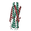
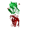
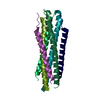
| Title | Respiratory syncytial virus fusion glycoprotein N-terminal heptad repeat domain+VIQKI I456F | |||||||||
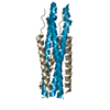
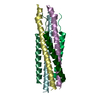
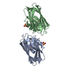
 Components Components | (Fusion glycoprotein F0) x 2 | |||||||||
 Keywords Keywords | ANTIVIRAL PROTEIN / Fusion protein / fusion inhibitor / six-helix bundle | |||||||||
| Function / homology |  Function and homology information Function and homology informationsymbiont-mediated induction of syncytium formation / Translation of respiratory syncytial virus mRNAs / RSV-host interactions / Assembly and release of respiratory syncytial virus (RSV) virions / Maturation of hRSV A proteins / Respiratory syncytial virus (RSV) attachment and entry / host cell Golgi membrane / entry receptor-mediated virion attachment to host cell / fusion of virus membrane with host plasma membrane / viral envelope ...symbiont-mediated induction of syncytium formation / Translation of respiratory syncytial virus mRNAs / RSV-host interactions / Assembly and release of respiratory syncytial virus (RSV) virions / Maturation of hRSV A proteins / Respiratory syncytial virus (RSV) attachment and entry / host cell Golgi membrane / entry receptor-mediated virion attachment to host cell / fusion of virus membrane with host plasma membrane / viral envelope / symbiont entry into host cell / host cell plasma membrane / virion membrane / identical protein binding / plasma membrane Similarity search - Function | |||||||||
| Biological species |  Human respiratory syncytial virus AHuman respirovirus 3 Human respiratory syncytial virus AHuman respirovirus 3 | |||||||||
| Method |  X-RAY DIFFRACTION / SYNCHROTRON / MOLECULAR REPLACEMENT / Resolution: 1.45 Å X-RAY DIFFRACTION / SYNCHROTRON / MOLECULAR REPLACEMENT / Resolution: 1.45 Å | |||||||||
 Authors Authors | Outlaw, V.K. / Gellman, S.H. | |||||||||
| Funding support |  United States, 2items United States, 2items
| |||||||||
 Citation Citation | Journal: J.Am.Chem.Soc. / Year: 2020 Title: Structure-Guided Improvement of a Dual HPIV3/RSV Fusion Inhibitor. Authors: Outlaw, V.K. / Lemke, J.T. / Zhu, Y. / Gellman, S.H. / Porotto, M. / Moscona, A. | |||||||||
| History |
|
- Structure visualization
| Structure viewer | Molecule: MolmilJmol/JSmol |
|---|
- Downloads & links
-Download
| PDBx/mmCIF format | 6oj7.cif.gz | 70.5 KB | Display | PDBx/mmCIF format |
|---|---|---|---|---|
| PDB format | pdb6oj7.ent.gz | 47.2 KB | Display | PDB format |
| PDBx/mmJSON format | 6oj7.json.gz | Tree view | PDBx/mmJSON format | |
| Others |  Other downloads Other downloads |
-Validation report
| Arichive directory | https://data.pdbj.org/pub/pdb/validation_reports/oj/6oj7ftp://data.pdbj.org/pub/pdb/validation_reports/oj/6oj7 | HTTPS FTP |
|---|
-Related structure data
| Related structure data | 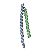 6o40C 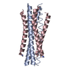 6ntxS S: Starting model for refinement C: citing same article ( |
|---|---|
| Similar structure data |


-Links
 PDBj
PDBj

- Assembly
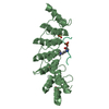
| Deposited unit | 
| ||||||||||||
|---|---|---|---|---|---|---|---|---|---|---|---|---|---|
| 1 | 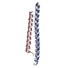
| ||||||||||||
| Unit cell |
|
-Components
| #1: Protein | Mass: 5564.542 Da / Num. of mol.: 1 / Fragment: N-terminal / Source method: obtained synthetically Details: Compound details: This compound is derived from the the RSV-A2 fusion glycoprotein N-terminal heptad repeat domain residues 158-208. It is acetylated at the N-terminus and amidated at the C-terminus. Source: (synth.) Human respiratory syncytial virus A / References: UniProt: A0A1U8ZTH8, UniProt: P03420*PLUS |
|---|---|
| #2: Protein/peptide | Mass: 4228.911 Da / Num. of mol.: 1 / Fragment: UNP residues 449-484 / Mutation: I456F, E459V, A463I, D466Q, Q479K, K480I / Source method: obtained synthetically Details: VIQKI I454F is a synthetic peptide derived from residues 449-484 of the HPIV3 fusion glycoprotein C-terminal heptad repeat domain with substitutions I454F, I456F, E459V, A463I, D466Q, Q479K, ...Details: VIQKI I454F is a synthetic peptide derived from residues 449-484 of the HPIV3 fusion glycoprotein C-terminal heptad repeat domain with substitutions I454F, I456F, E459V, A463I, D466Q, Q479K, and K480I. It is acetylated at the N-terminus and amidated at the C-terminus. Source: (synth.) Human respirovirus 3 / References: UniProt: A0A1X9QNS5, UniProt: P06828*PLUS |
| #3: Water | ChemComp-HOH /  Mass: 18.015 Da / Num. of mol.: 65 / Source method: isolated from a natural source / Formula: H2O Mass: 18.015 Da / Num. of mol.: 65 / Source method: isolated from a natural source / Formula: H2O |
| Has protein modification | Y |
-Experimental details
-Experiment
| Experiment | Method: X-RAY DIFFRACTION / Number of used crystals: 1 |
|---|
- Sample preparation
| Crystal | Density Matthews: 2.19 Å3/Da / Density % sol: 43.87 % |
|---|---|
| Crystal grow | Temperature: 298 K / Method: vapor diffusion, hanging drop / pH: 7.5 Details: 30 mM NaF, 30 mM NaBr, 30 mM NaI, 12.5% (v/v) 2-methyl-2,4-pentanediol, 12.5% (v/v) PEG 1000, 12.5% (w/v) PEG 3350 in 100 mM NaHEPES/MOPS buffer (pH 7.5) |
-Data collection
| Diffraction | Mean temperature: 100 K / Serial crystal experiment: N |
|---|---|
| Diffraction source | Source: SYNCHROTRON / Site: APS / Beamline: 21-ID-D / Wavelength: 1.127231 Å |
| Detector | Type: DECTRIS EIGER X 9M / Detector: PIXEL / Date: Jul 25, 2018 |
| Radiation | Protocol: SINGLE WAVELENGTH / Monochromatic (M) / Laue (L): M / Scattering type: x-ray |
| Radiation wavelength | Wavelength: 1.127231 Å / Relative weight: 1 |
| Reflection | Resolution: 1.45→26.96 Å / Num. obs: 13875 / % possible obs: 98.55 % / Redundancy: 9.86 % / Biso Wilson estimate: 19.28 Å2 / CC1/2: 0.995 / Rmerge(I) obs: 0.114 / Rpim(I) all: 0.0374 / Rrim(I) all: 0.12 / Net I/σ(I): 9.19 |
| Reflection shell | Resolution: 1.45→1.502 Å / Redundancy: 6.3 % / Rmerge(I) obs: 0.85 / Mean I/σ(I) obs: 1.51 / Num. unique obs: 1262 / CC1/2: 0.399 / Rpim(I) all: 0.366 / Rrim(I) all: 0.929 / % possible all: 89.01 |
- Processing
| Software |
| |||||||||||||||||||||||||||||||||||||||||||||||||||||||||||||||||||||||||||||
|---|---|---|---|---|---|---|---|---|---|---|---|---|---|---|---|---|---|---|---|---|---|---|---|---|---|---|---|---|---|---|---|---|---|---|---|---|---|---|---|---|---|---|---|---|---|---|---|---|---|---|---|---|---|---|---|---|---|---|---|---|---|---|---|---|---|---|---|---|---|---|---|---|---|---|---|---|---|---|
| Refinement | Method to determine structure: MOLECULAR REPLACEMENT Starting model: 6NTX Resolution: 1.45→26.96 Å / SU ML: 0.1803 / Cross valid method: FREE R-VALUE / σ(F): 1.97 / Phase error: 22.4085
| |||||||||||||||||||||||||||||||||||||||||||||||||||||||||||||||||||||||||||||
| Solvent computation | Shrinkage radii: 0.9 Å / VDW probe radii: 1.11 Å | |||||||||||||||||||||||||||||||||||||||||||||||||||||||||||||||||||||||||||||
| Displacement parameters | Biso mean: 40.05 Å2 | |||||||||||||||||||||||||||||||||||||||||||||||||||||||||||||||||||||||||||||
| Refinement step | Cycle: LAST / Resolution: 1.45→26.96 Å
| |||||||||||||||||||||||||||||||||||||||||||||||||||||||||||||||||||||||||||||
| Refine LS restraints |
| |||||||||||||||||||||||||||||||||||||||||||||||||||||||||||||||||||||||||||||
| LS refinement shell |
| |||||||||||||||||||||||||||||||||||||||||||||||||||||||||||||||||||||||||||||
| Refinement TLS params. | Method: refined / Origin x: 1.17329727507 Å / Origin y: 6.92552153942 Å / Origin z: 10.4728440421 Å
| |||||||||||||||||||||||||||||||||||||||||||||||||||||||||||||||||||||||||||||
| Refinement TLS group | Selection details: all |
