 Movie
Movie Controller

[English] 日本語
 Yorodumi
- PDB-1g3p: CRYSTAL STRUCTURE OF THE N-TERMINAL DOMAINS OF BACTERIOPHAGE MINO... -
+ Open data

- Basic information
| Entry | Database: PDB / ID: 1g3p | ||||||
|---|---|---|---|---|---|---|---|
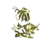
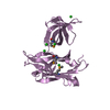
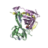
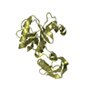
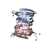
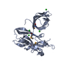
| Title | CRYSTAL STRUCTURE OF THE N-TERMINAL DOMAINS OF BACTERIOPHAGE MINOR COAT PROTEIN G3P | ||||||
 Components Components | MINOR COAT PROTEIN | ||||||
 Keywords Keywords | VIRAL PROTEIN / MINOR COAT PROTEIN / FILAMENTOUS BACTERIOPHAGE / PHAGE DISPLAY / SELECTIVELY INFECTIVE PHAGES | ||||||
| Function / homology |  Function and homology information Function and homology informationviral extrusion / virion attachment to host cell pilus / adhesion receptor-mediated virion attachment to host cell / host cell membrane / viral capsid / entry receptor-mediated virion attachment to host cell Similarity search - Function | ||||||
| Biological species |  Enterobacteria phage M13 (virus) Enterobacteria phage M13 (virus) | ||||||
| Method |  X-RAY DIFFRACTION / SYNCHROTRON / SIRAS (USING MAD X-RAY DATA) / Resolution: 1.46 Å X-RAY DIFFRACTION / SYNCHROTRON / SIRAS (USING MAD X-RAY DATA) / Resolution: 1.46 Å | ||||||
 Authors Authors | Lubkowski, J. / Hennecke, F. / Pluckthun, A. / Wlodawer, A. | ||||||
 Citation Citation | Journal: Nat.Struct.Biol. / Year: 1998 Title: The structural basis of phage display elucidated by the crystal structure of the N-terminal domains of g3p. Authors: Lubkowski, J. / Hennecke, F. / Pluckthun, A. / Wlodawer, A. #1: Journal: Nat.Med. (N.Y.) / Year: 1997Title: Selectively Infective Phage (Sip) Technology: A Novel Method for in Vivo Selection of Interacting Protein-Ligand Pairs Authors: Spada, S. / Pluckthun, A. #2: Journal: Cell(Cambridge,Mass.) / Year: 1997Title: The C-Terminal Domain of Tola is the Coreceptor for Filamentous Phage Infection of E. Coli Authors: Riechmann, L. / Holliger, P. #3: Journal: Structure / Year: 1997Title: A Conserved Infection Pathway for Filamentous Bacteriophages is Suggested by the Structure of the Membrane Penetration Domain of the Minor Coat Protein G3P from Phage Fd Authors: Holliger, P. / Riechmann, L. | ||||||
| History |
|
- Structure visualization
| Structure viewer | Molecule: MolmilJmol/JSmol |
|---|
- Downloads & links
-Download
| PDBx/mmCIF format | 1g3p.cif.gz | 61 KB | Display | PDBx/mmCIF format |
|---|---|---|---|---|
| PDB format | pdb1g3p.ent.gz | 43.9 KB | Display | PDB format |
| PDBx/mmJSON format | 1g3p.json.gz | Tree view | PDBx/mmJSON format | |
| Others |  Other downloads Other downloads |
-Validation report
| Arichive directory | https://data.pdbj.org/pub/pdb/validation_reports/g3/1g3pftp://data.pdbj.org/pub/pdb/validation_reports/g3/1g3p | HTTPS FTP |
|---|
-Related structure data
| Similar structure data |
|---|


-Links
 PDBj
PDBj- Assembly
| Deposited unit | 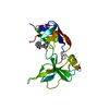
| |||||||||
|---|---|---|---|---|---|---|---|---|---|---|
| 1 |
| |||||||||
| Unit cell |
| |||||||||
| Components on special symmetry positions |
|
-Components
| #1: Protein | Mass: 23626.484 Da / Num. of mol.: 1 / Fragment: TWO N-TERMINAL DOMAINS, N1 AND N2 Source method: isolated from a genetically manipulated source Source: (gene. exp.) Enterobacteria phage M13 (virus) / Genus: Inovirus / Strain: M13MP18 / Cell line: BL21 (DE3) FOR / Gene: 3 / Plasmid: BL21Cell line (production host): BL21 (DE3), FOR SELENOMETHIONINE-VARIANT DL41 (DE3) Production host:  |
|---|---|
| #2: Chemical | ChemComp-SO4 /   Mass: 96.063 Da / Num. of mol.: 1 / Source method: obtained synthetically / Formula: SO4 Mass: 96.063 Da / Num. of mol.: 1 / Source method: obtained synthetically / Formula: SO4 |
| #3: Water | ChemComp-HOH /  Mass: 18.015 Da / Num. of mol.: 312 / Source method: isolated from a natural source / Formula: H2O Mass: 18.015 Da / Num. of mol.: 312 / Source method: isolated from a natural source / Formula: H2O |
| Compound details | MINOR COAT PROTEIN FROM GENE 3 OF FILAMENTOUS BACTERIOPHAGE M13. THE PHAGE COAT PROTEIN (G3P) ...MINOR COAT PROTEIN FROM GENE 3 OF FILAMENTOU |
| Has protein modification | Y |
| Nonpolymer details | TRO 21 IS THE OXIDIZED TRP. TRO CARRIES MOST LIKELY AN OXINDOLE SIDE CHAIN (1,3-DIHYDRO-INDOLE-2- ...TRO 21 IS THE OXIDIZED TRP. TRO CARRIES MOST LIKELY AN OXINDOLE SIDE CHAIN (1,3-DIHYDRO-INDOLE-2-ONE). THE OXYGEN ATOM IS BOUND (LIKELY VIA A DOUBLE BOND) TO THE CD1 CARBON. ADDITIONAL |
| Sequence details | THE SEQUENCE DIFFERENCES ARE NATURALLY OCCURRING VARIATIONS WHICH DO NOT AFFECT THE FUNCTIONALITY ...THE SEQUENCE DIFFERENCE |
-Experimental details
-Experiment
| Experiment | Method: X-RAY DIFFRACTION / Number of used crystals: 1 |
|---|
- Sample preparation
| Crystal | Density Matthews: 2.34 Å3/Da / Density % sol: 40 % | ||||||||||||||||||||||||||||||
|---|---|---|---|---|---|---|---|---|---|---|---|---|---|---|---|---|---|---|---|---|---|---|---|---|---|---|---|---|---|---|---|
| Crystal grow | Method: vapor diffusion, hanging drop / pH: 6.5 Details: EQUAL VOLUMES OF THE PROTEIN SOLUTION (10 MG/ML) BUFFERED WITH 50 MM PIPES PH 6.5 AND THE PRECIPITANT (30% PEG 4000, 0.2 M AMMONIUM SULFATE, 2 MM DTT) WERE MIXED AND EQUILIBRATED (IN THE ...Details: EQUAL VOLUMES OF THE PROTEIN SOLUTION (10 MG/ML) BUFFERED WITH 50 MM PIPES PH 6.5 AND THE PRECIPITANT (30% PEG 4000, 0.2 M AMMONIUM SULFATE, 2 MM DTT) WERE MIXED AND EQUILIBRATED (IN THE HANGING DROP SETUP) AGAINST THE PRECIPITANT., vapor diffusion - hanging drop | ||||||||||||||||||||||||||||||
| Crystal grow | *PLUS Method: unknown | ||||||||||||||||||||||||||||||
| Components of the solutions | *PLUS
|
-Data collection
| Diffraction | Mean temperature: 100 K |
|---|---|
| Diffraction source | Source: SYNCHROTRON / Site: NSLS  / Beamline: X9B / Wavelength: 0.98 / Beamline: X9B / Wavelength: 0.98 |
| Detector | Type: MAR scanner 345 mm plate / Detector: IMAGE PLATE / Date: Sep 1, 1997 / Details: COLLIMATOR |
| Radiation | Monochromator: SI(111) / Monochromatic (M) / Laue (L): M / Scattering type: x-ray |
| Radiation wavelength | Wavelength: 0.98 Å / Relative weight: 1 |
| Reflection | Resolution: 1.46→22 Å / Num. obs: 37511 / % possible obs: 99.8 % / Observed criterion σ(I): -3 / Redundancy: 7.53 % / Rsym value: 0.034 / Net I/σ(I): 27 |
| Reflection shell | Resolution: 1.46→1.51 Å / Redundancy: 5.5 % / Mean I/σ(I) obs: 8.5 / Rsym value: 0.11 / % possible all: 99.2 |
| Reflection | *PLUS Num. measured all: 282603 / Rmerge(I) obs: 0.034 |
| Reflection shell | *PLUS % possible obs: 99.2 % / Rmerge(I) obs: 0.11 |
- Processing
| Software |
| ||||||||||||||||||||||||||||||||||||||||||||||||||||||||||||||||||||||||||||||||
|---|---|---|---|---|---|---|---|---|---|---|---|---|---|---|---|---|---|---|---|---|---|---|---|---|---|---|---|---|---|---|---|---|---|---|---|---|---|---|---|---|---|---|---|---|---|---|---|---|---|---|---|---|---|---|---|---|---|---|---|---|---|---|---|---|---|---|---|---|---|---|---|---|---|---|---|---|---|---|---|---|---|
| Refinement | Method to determine structure: SIRAS (USING MAD X-RAY DATA) / Resolution: 1.46→8 Å / Rfactor Rfree error: 0.0037 / Data cutoff high absF: 10000000 / Data cutoff low absF: 0.001 / Isotropic thermal model: RESTRAINED / Cross valid method: THROUGHOUT / σ(F): 2 Details: SEVEN RESIDUES WERE REFINED IN TWO ALTERNATE CONFORMATIONS EACH.
| ||||||||||||||||||||||||||||||||||||||||||||||||||||||||||||||||||||||||||||||||
| Displacement parameters | Biso mean: 16.1 Å2 | ||||||||||||||||||||||||||||||||||||||||||||||||||||||||||||||||||||||||||||||||
| Refinement step | Cycle: LAST / Resolution: 1.46→8 Å
| ||||||||||||||||||||||||||||||||||||||||||||||||||||||||||||||||||||||||||||||||
| Refine LS restraints |
| ||||||||||||||||||||||||||||||||||||||||||||||||||||||||||||||||||||||||||||||||
| LS refinement shell | Resolution: 1.46→1.53 Å / Rfactor Rfree error: 0.011 / Total num. of bins used: 8
| ||||||||||||||||||||||||||||||||||||||||||||||||||||||||||||||||||||||||||||||||
| Xplor file |
| ||||||||||||||||||||||||||||||||||||||||||||||||||||||||||||||||||||||||||||||||
| Software | *PLUS Name: X-PLOR / Version: 3.1 / Classification: refinement | ||||||||||||||||||||||||||||||||||||||||||||||||||||||||||||||||||||||||||||||||
| Refine LS restraints | *PLUS
|
