 Movie
Movie Controller

[English] 日本語
 Yorodumi
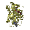
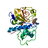
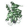
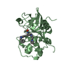
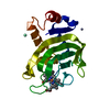
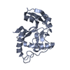
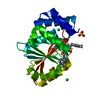
- PDB-4ekf: Structure of the Inactive Adenovirus Proteinase at 0.98 Angstrom ... -
+ Open data

- Basic information
| Entry | Database: PDB / ID: 4ekf | ||||||
|---|---|---|---|---|---|---|---|
| Title | Structure of the Inactive Adenovirus Proteinase at 0.98 Angstrom Resolution | ||||||
 Components Components | Adenain | ||||||
 Keywords Keywords | HYDROLASE / alpha and beta protein (a+b) | ||||||
| Function / homology |  Function and homology information Function and homology informationadenain / cysteine-type peptidase activity / virion component / cysteine-type endopeptidase activity / host cell nucleus / proteolysis / DNA binding Similarity search - Function | ||||||
| Biological species |   Human adenovirus 2 Human adenovirus 2 | ||||||
| Method |  X-RAY DIFFRACTION / SYNCHROTRON / MOLECULAR REPLACEMENT / molecular replacement / Resolution: 0.98 Å X-RAY DIFFRACTION / SYNCHROTRON / MOLECULAR REPLACEMENT / molecular replacement / Resolution: 0.98 Å | ||||||
 Authors Authors | Baniecki, M.L. / McGrath, W.J. / Mangel, W.F. | ||||||
 Citation Citation | Journal: J.Biol.Chem. / Year: 2013 Title: Regulation of a Viral Proteinase by a Peptide and DNA in One-dimensional Space: III. ATOMIC RESOLUTION STRUCTURE OF THE NASCENT FORM OF THE ADENOVIRUS PROTEINASE. Authors: Baniecki, M.L. / McGrath, W.J. / Mangel, W.F. #1: Journal: J.Biol.Chem. / Year: 2012 Title: Regulation of a viral proteinase by a peptide and DNA in one-dimensional space. I. binding to DNA and to hexon of the precursor to protein VI, pVI, of human adenovirus. Authors: Graziano, V. / McGrath, W.J. / Suomalainen, M. / Greber, U.F. / Freimuth, P. / Blainey, P.C. / Luo, G. / Xie, X.S. / Mangel, W.F. #2: Journal: J.Biol.Chem. / Year: 2012 Title: Regulation of a viral proteinase by a peptide and DNA in one-dimensional space. II. adenovirus proteinase is activated in an unusual one-dimensional biochemical reaction. Authors: Graziano, V. / Luo, G. / Blainey, P.C. / Perez-Berna, A.J. / McGrath, W.J. / Flint, S.J. / San Martin, C. / Xie, X.S. / Mangel, W.F. #3: Journal: J.Biol.Chem. / Year: 2012 Title: Regulation of a viral proteinase by a peptide and DNA in one-dimensional space. IV. viral proteinase slides along DNA to locate and process its substrates. Authors: Blainey, P.C. / Graziano, V. / Perez-Berna, A.J. / McGrath, W.J. / Flint, S.J. / San Martin, C. / Xie, X.S. / Mangel, W.F. #4: Journal: Acta Crystallogr.,Sect.D / Year: 2002 Title: Adenovirus proteinase: crystallization and preliminary X-ray diffraction studies to atomic resolution. Authors: Baniecki, M.L. / McGrath, W.J. / Dauter, Z. / Mangel, W.F. #5: Journal: Embo J. / Year: 1996Title: Crystal structure of the human adenovirus proteinase with its 11 amino acid cofactor. Authors: Ding, J. / McGrath, W.J. / Sweet, R.M. / Mangel, W.F. #6: Journal: Biochim.Biophys.Acta / Year: 2003Title: Crystallographic structure at 1.6-A resolution of the human adenovirus proteinase in a covalent complex with its 11-amino-acid peptide cofactor: insights on a new fold. Authors: McGrath, W.J. / Ding, J. / Didwania, A. / Sweet, R.M. / Mangel, W.F. #7: Journal: Nature / Year: 1993 Title: Viral DNA and a viral peptide can act as cofactors of adenovirus virion proteinase activity. Authors: Mangel, W.F. / McGrath, W.J. / Toledo, D.L. / Anderson, C.W. | ||||||
| History |
|
- Structure visualization
| Structure viewer | Molecule: MolmilJmol/JSmol |
|---|
- Downloads & links
-Download
| PDBx/mmCIF format | 4ekf.cif.gz | 132.8 KB | Display | PDBx/mmCIF format |
|---|---|---|---|---|
| PDB format | pdb4ekf.ent.gz | 103.6 KB | Display | PDB format |
| PDBx/mmJSON format | 4ekf.json.gz | Tree view | PDBx/mmJSON format | |
| Others |  Other downloads Other downloads |
-Validation report
| Arichive directory | https://data.pdbj.org/pub/pdb/validation_reports/ek/4ekfftp://data.pdbj.org/pub/pdb/validation_reports/ek/4ekf | HTTPS FTP |
|---|
-Related structure data
| Related structure data | 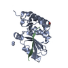 1nlnS S: Starting model for refinement |
|---|---|
| Similar structure data |


-Links
 PDBj
PDBj- Assembly
| Deposited unit | 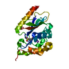
| ||||||||
|---|---|---|---|---|---|---|---|---|---|
| 1 |
| ||||||||
| Unit cell |
|
-Components
| #1: Protein | Mass: 23146.348 Da / Num. of mol.: 1 Source method: isolated from a genetically manipulated source Source: (gene. exp.) Human adenovirus 2 / Strain: serotype 2 / Gene: L3-23K / Plasmid: pET 13a / Production host:  |
|---|---|
| #2: Chemical | ChemComp-NA /   Mass: 22.990 Da / Num. of mol.: 1 / Source method: obtained synthetically / Formula: Na Mass: 22.990 Da / Num. of mol.: 1 / Source method: obtained synthetically / Formula: Na |
| #3: Water | ChemComp-HOH /  Mass: 18.015 Da / Num. of mol.: 258 / Source method: isolated from a natural source / Formula: H2O Mass: 18.015 Da / Num. of mol.: 258 / Source method: isolated from a natural source / Formula: H2O |
| Has protein modification | Y |
-Experimental details
-Experiment
| Experiment | Method: X-RAY DIFFRACTION / Number of used crystals: 1 |
|---|
- Sample preparation
| Crystal | Density Matthews: 1.75 Å3/Da / Density % sol: 29.2 % |
|---|---|
| Crystal grow | Temperature: 293 K / pH: 5.6 Details: 0.3M sodium citrate, pH 5.6, 0.8 M sodium acetate, 5 mM DTT, VAPOR DIFFUSION, HANGING DROP, temperature 293K |
-Data collection
| Diffraction | Mean temperature: 100 K |
|---|---|
| Diffraction source | Source: SYNCHROTRON / Site: NSLS  / Beamline: X25 / Wavelength: 0.92 / Beamline: X25 / Wavelength: 0.92 |
| Detector | Type: BRANDEIS 4K / Detector: CCD / Date: Jul 28, 2001 |
| Radiation | Protocol: SINGLE WAVELENGTH / Monochromatic (M) / Laue (L): M / Scattering type: x-ray |
| Radiation wavelength | Wavelength: 0.92 Å / Relative weight: 1 |
| Reflection | Resolution: 0.98→30 Å / Num. obs: 92231 / % possible obs: 99.1 % / Redundancy: 4.9 % / Biso Wilson estimate: 8.59 Å2 / Rmerge(I) obs: 0.057 / Net I/σ(I): 25.4 |
| Reflection shell | Resolution: 0.98→1 Å / Redundancy: 2.2 % / Rmerge(I) obs: 0.594 / % possible all: 97.1 |
-Phasing
| Phasing | Method: molecular replacement |
|---|
- Processing
| Software |
| ||||||||||||||||||||||||||||||||||||
|---|---|---|---|---|---|---|---|---|---|---|---|---|---|---|---|---|---|---|---|---|---|---|---|---|---|---|---|---|---|---|---|---|---|---|---|---|---|
| Refinement | Method to determine structure: MOLECULAR REPLACEMENT Starting model: PDB ENTRY 1NLN CHAIN A Resolution: 0.98→20 Å / Num. parameters: 16135 / Num. restraintsaints: 19747 / Occupancy max: 1 / Occupancy min: 0 / Cross valid method: FREE R / σ(F): 0 / Stereochemistry target values: ENGH AND HUBER Details: ANISOTROPIC REFINEMENT REDUCED FREE R (NO CUTOFF) BY ?
| ||||||||||||||||||||||||||||||||||||
| Displacement parameters | Biso mean: 17.7814 Å2 | ||||||||||||||||||||||||||||||||||||
| Refine analyze | Luzzati coordinate error obs: 0.127 Å / Num. disordered residues: 12 / Occupancy sum hydrogen: 1406 / Occupancy sum non hydrogen: 1732.98 | ||||||||||||||||||||||||||||||||||||
| Refinement step | Cycle: LAST / Resolution: 0.98→20 Å
| ||||||||||||||||||||||||||||||||||||
| Refine LS restraints |
|
