 Movie
Movie Controller

[English] 日本語
 Yorodumi
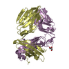
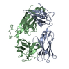
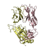
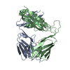
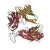
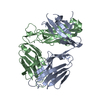
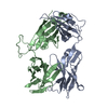
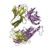
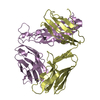
- PDB-4ei6: Structure of XV19 Valpha1-Vbeta16 Type-II Natural Killer T cell r... -
+ Open data

- Basic information
| Entry | Database: PDB / ID: 4ei6 | ||||||
|---|---|---|---|---|---|---|---|
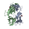
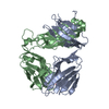
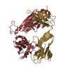
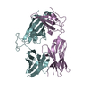
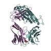
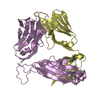
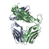
| Title | Structure of XV19 Valpha1-Vbeta16 Type-II Natural Killer T cell receptor | ||||||
 Components Components |
| ||||||
 Keywords Keywords | IMMUNE SYSTEM / Natural Killer T cell receptor | ||||||
| Function / homology | Immunoglobulins / Immunoglobulin-like / Sandwich / Mainly Beta Function and homology information Function and homology information | ||||||
| Biological species |   Homo sapiens (human) Homo sapiens (human) | ||||||
| Method |  X-RAY DIFFRACTION / SYNCHROTRON / MOLECULAR REPLACEMENT / Resolution: 1.6 Å X-RAY DIFFRACTION / SYNCHROTRON / MOLECULAR REPLACEMENT / Resolution: 1.6 Å | ||||||
 Authors Authors | Patel, O. / Rossjohn, J. | ||||||
 Citation Citation | Journal: Nat.Immunol. / Year: 2012 Title: Recognition of CD1d-sulfatide mediated by a type II natural killer T cell antigen receptor. Authors: Patel, O. / Pellicci, D.G. / Gras, S. / Sandoval-Romero, M.L. / Uldrich, A.P. / Mallevaey, T. / Clarke, A.J. / Le Nours, J. / Theodossis, A. / Cardell, S.L. / Gapin, L. / Godfrey, D.I. / Rossjohn, J. | ||||||
| History |
|
- Structure visualization
| Structure viewer | Molecule: MolmilJmol/JSmol |
|---|
- Downloads & links
-Download
| PDBx/mmCIF format | 4ei6.cif.gz | 201 KB | Display | PDBx/mmCIF format |
|---|---|---|---|---|
| PDB format | pdb4ei6.ent.gz | 158.2 KB | Display | PDB format |
| PDBx/mmJSON format | 4ei6.json.gz | Tree view | PDBx/mmJSON format | |
| Others |  Other downloads Other downloads |
-Validation report
| Summary document | 4ei6_validation.pdf.gz | 446.6 KB | Display | wwPDB validaton report |
|---|---|---|---|---|
| Full document | 4ei6_full_validation.pdf.gz | 453.9 KB | Display | |
| Data in XML | 4ei6_validation.xml.gz | 40.4 KB | Display | |
| Data in CIF | 4ei6_validation.cif.gz | 60.5 KB | Display | |
| Arichive directory | https://data.pdbj.org/pub/pdb/validation_reports/ei/4ei6ftp://data.pdbj.org/pub/pdb/validation_reports/ei/4ei6 | HTTPS FTP |
-Related structure data
| Related structure data |  4ei5C 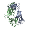 2bnuS C: citing same article ( S: Starting model for refinement |
|---|---|
| Similar structure data |


-Links
 PDBj
PDBj- Assembly
| Deposited unit | 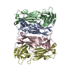
| ||||||||
|---|---|---|---|---|---|---|---|---|---|
| 1 | 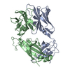
| ||||||||
| 2 | 
| ||||||||
| Unit cell |
|
-Components
| #1: Protein | Mass: 23209.518 Da / Num. of mol.: 2 / Fragment: extracellular domain (SEE REMARK 999) Source method: isolated from a genetically manipulated source Source: (gene. exp.) Mus musculus, Homo sapiens / Plasmid: pET30b / Production host:  #2: Protein | Mass: 28080.617 Da / Num. of mol.: 2 / Fragment: extracellular domain (SEE REMARK 999) Source method: isolated from a genetically manipulated source Source: (gene. exp.) Mus musculus, Homo sapiens / Plasmid: pET30b / Production host: #3: Water | ChemComp-HOH / |  Mass: 18.015 Da / Num. of mol.: 816 / Source method: isolated from a natural source / Formula: H2O Mass: 18.015 Da / Num. of mol.: 816 / Source method: isolated from a natural source / Formula: H2OHas protein modification | Y | Sequence details | CHAINS A AND C ARE CHIMERAS COMPRISING THE MOUSE VARIABLE DOMAIN (RESIDUES 1-116) AND HUMAN ...CHAINS A AND C ARE CHIMERAS COMPRISING | |
|---|
-Experimental details
-Experiment
| Experiment | Method: X-RAY DIFFRACTION / Number of used crystals: 1 |
|---|
- Sample preparation
| Crystal | Density Matthews: 2.43 Å3/Da / Density % sol: 49.41 % |
|---|---|
| Crystal grow | Temperature: 293 K / Method: vapor diffusion, hanging drop / pH: 6 Details: 0.1 M sodium malonate/imidazole/boric acid buffer, 25% PEG3350, pH 6, VAPOR DIFFUSION, HANGING DROP, temperature 293K |
-Data collection
| Diffraction | Mean temperature: 100 K |
|---|---|
| Diffraction source | Source: SYNCHROTRON / Site: Australian Synchrotron  / Beamline: MX2 / Wavelength: 0.95453 Å / Beamline: MX2 / Wavelength: 0.95453 Å |
| Detector | Type: ADSC QUANTUM 315r / Detector: CCD / Date: Feb 18, 2011 |
| Radiation | Monochromator: Si(111) / Protocol: SINGLE WAVELENGTH / Monochromatic (M) / Laue (L): M / Scattering type: x-ray |
| Radiation wavelength | Wavelength: 0.95453 Å / Relative weight: 1 |
| Reflection | Resolution: 1.6→50 Å / Num. all: 132448 / Num. obs: 132448 / % possible obs: 100 % / Observed criterion σ(F): 0 / Observed criterion σ(I): 0 / Redundancy: 7.9 % / Biso Wilson estimate: 21.64 Å2 / Rmerge(I) obs: 0.079 / Net I/σ(I): 14.2 |
| Reflection shell | Resolution: 1.6→1.69 Å / Redundancy: 8.1 % / Rmerge(I) obs: 1.001 / Mean I/σ(I) obs: 1.9 / Num. unique all: 19137 / % possible all: 100 |
- Processing
| Software |
| |||||||||||||||||||||||||
|---|---|---|---|---|---|---|---|---|---|---|---|---|---|---|---|---|---|---|---|---|---|---|---|---|---|---|
| Refinement | Method to determine structure: MOLECULAR REPLACEMENT Starting model: PDB ENTRY 2BNU Resolution: 1.6→44.45 Å / SU ML: 0.23 / Cross valid method: THROUGHOUT / σ(F): 0 / Phase error: 23.11 / Stereochemistry target values: ML
| |||||||||||||||||||||||||
| Solvent computation | Shrinkage radii: 0.95 Å / VDW probe radii: 1.2 Å / Solvent model: FLAT BULK SOLVENT MODEL / Bsol: 43.348 Å2 / ksol: 0.351 e/Å3 | |||||||||||||||||||||||||
| Displacement parameters |
| |||||||||||||||||||||||||
| Refinement step | Cycle: LAST / Resolution: 1.6→44.45 Å
| |||||||||||||||||||||||||
| Refine LS restraints |
| |||||||||||||||||||||||||
| LS refinement shell | Resolution: 1.6→1.69 Å
|
