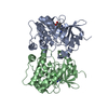
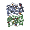
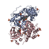
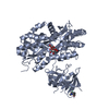
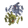
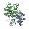
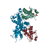
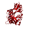
登録情報 データベース : PDB / ID : 4edqタイトル MBP-fusion protein of myosin-binding protein c residues 149-269 Maltose-binding periplasmic protein,Myosin-binding protein C, cardiac-type chimeric protein キーワード / / / / 機能・相同性 分子機能 ドメイン・相同性 構成要素
/ / / / / / / / / / / / / / / / / / / / / / / / / / / / / / / / / / / / / / / / / / / / / / / / / / / / / / / / / / / / / / / / / / / / / / / / / / / / / 生物種 Escherichia coli (大腸菌)Mus musculus (ハツカネズミ)手法 / / / 解像度 : 1.641 Å データ登録者 Rebowski, G. / Boczkowska, M. / Dominguez, R. ジャーナル : To be Published タイトル : MBP-fusion protein of myosin-binding protein c residues 149-269著者 : Rebowski, G. / Boczkowska, M. / Dominguez, R. 履歴 登録 2012年3月27日 登録サイト / 処理サイト 改定 1.0 2013年3月20日 Provider / タイプ 改定 1.1 2017年7月26日 Group / Source and taxonomy / カテゴリ / entity_src_gen / Item 改定 2.0 2020年7月29日 Group Advisory / Atomic model ... Advisory / Atomic model / Data collection / Database references / Derived calculations / Non-polymer description / Structure summary カテゴリ atom_site / atom_site_anisotrop ... atom_site / atom_site_anisotrop / chem_comp / database_PDB_caveat / entity / entity_name_com / pdbx_branch_scheme / pdbx_chem_comp_identifier / pdbx_entity_branch / pdbx_entity_branch_descriptor / pdbx_entity_branch_link / pdbx_entity_branch_list / pdbx_entity_nonpoly / pdbx_molecule_features / pdbx_nonpoly_scheme / struct_conn / struct_ref_seq_dif / struct_site / struct_site_gen Item _atom_site.B_iso_or_equiv / _atom_site.Cartn_x ... _atom_site.B_iso_or_equiv / _atom_site.Cartn_x / _atom_site.Cartn_y / _atom_site.Cartn_z / _atom_site.auth_asym_id / _atom_site.auth_atom_id / _atom_site.auth_comp_id / _atom_site.auth_seq_id / _atom_site.label_atom_id / _atom_site.label_comp_id / _atom_site.type_symbol / _atom_site_anisotrop.id / _atom_site_anisotrop.pdbx_auth_asym_id / _atom_site_anisotrop.pdbx_auth_atom_id / _atom_site_anisotrop.pdbx_auth_comp_id / _atom_site_anisotrop.pdbx_auth_seq_id / _atom_site_anisotrop.pdbx_label_atom_id / _atom_site_anisotrop.pdbx_label_comp_id / _chem_comp.formula / _chem_comp.formula_weight / _chem_comp.id / _chem_comp.mon_nstd_flag / _chem_comp.name / _chem_comp.pdbx_synonyms / _chem_comp.type / _entity.formula_weight / _entity.pdbx_description / _entity.src_method / _entity.type / _struct_ref_seq_dif.details 解説 / Provider / タイプ 改定 2.1 2023年9月13日 Group Data collection / Database references ... Data collection / Database references / Refinement description / Structure summary カテゴリ chem_comp / chem_comp_atom ... chem_comp / chem_comp_atom / chem_comp_bond / database_2 / pdbx_initial_refinement_model Item / _database_2.pdbx_DOI / _database_2.pdbx_database_accession
すべて表示 表示を減らす
 ムービー
ムービー コントローラー


 データを開く
 基本情報
基本情報 要素
要素 キーワード
キーワード 機能・相同性情報

 X線回折 /
  データ登録者
 引用
引用 構造の表示
構造の表示 ダウンロードとリンク
 その他のダウンロード
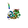


 PDBj
PDBj


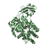
 集合体
集合体


 分子量: 18.015 Da / 分子数: 1300 / 由来タイプ: 天然 / 式: H2O
 試料調製
試料調製 / ビームライン: 17-ID / 波長: 0.9789 Å
 解析
解析