- PDB-3r13: Crystal structure of a Deoxyribose-phosphate aldolase (TM_1559) f... -
+
Open data
ID or keywords:
Loading...
-
Basic information
Entry
Database: PDB / ID: 3r13
Title
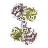
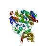
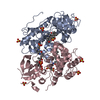
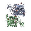
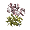
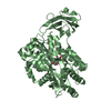
Crystal structure of a Deoxyribose-phosphate aldolase (TM_1559) from THERMOTOGA MARITIMA at 1.83 A resolution
Components
Deoxyribose-phosphate aldolase
Keywords
LYASE / TIM beta/alpha-barrel / Structural Genomics / Joint Center for Structural Genomics / JCSG / Protein Structure Initiative / PSI
Function / homology
Function and homology information
deoxyribose-phosphate aldolase / deoxyribose-phosphate aldolase activity / 2-deoxyribose 1-phosphate catabolic process / deoxyribonucleotide catabolic process / carbohydrate catabolic process / cytoplasm Similarity search - Function
Deoxyribose-phosphate aldolase type I / Deoxyribose-phosphate aldolase / DeoC/LacD family aldolase / DeoC/FbaB/LacD aldolase / DeoC/LacD family aldolase / Aldolase class I / Aldolase-type TIM barrel / TIM Barrel / Alpha-Beta Barrel / Alpha Beta Similarity search - Domain/homology
Resolution: 1.83→44.677 Å / Num. obs: 42195 / % possible obs: 100 % / Redundancy: 3.7 % / Rmerge(I) obs: 0.094 / Rsym value: 0.094 / Net I/σ(I): 8.2
Reflection shell
Diffraction-ID: 1
Resolution (Å)
Redundancy (%)
Rmerge(I) obs
Mean I/σ(I) obs
Num. measured all
Num. unique all
Rsym value
% possible all
1.83-1.93
3.7
0.552
2.4
23040
6174
0.552
100
1.93-2.05
3.8
0.4
3.3
21478
5725
0.4
100
2.05-2.19
3.8
0.27
4.7
20571
5482
0.27
100
2.19-2.36
3.8
0.2
6
19024
5059
0.2
100
2.36-2.59
3.8
0.151
7.4
17609
4672
0.151
100
2.59-2.89
3.8
0.108
9.7
16019
4252
0.108
100
2.89-3.34
3.8
0.075
12.4
14093
3754
0.075
100
3.34-4.09
3.7
0.05
16.9
11899
3186
0.05
100
4.09-5.79
3.7
0.039
20.2
9209
2486
0.039
100
5.79-44.677
3.6
0.034
21.6
5003
1405
0.034
99.7
-
Phasing
Phasing
Method: MAD
-
Processing
Software
Name
Version
Classification
NB
MolProbity
3beta29
modelbuilding
PDB_EXTRACT
3.1
dataextraction
SHELX
phasing
SHARP
phasing
SCALA
3.2.5
datascaling
REFMAC
5.5.0110
refinement
XDS
datareduction
XSCALE
datascaling
SHELXD
phasing
autoSHARP
phasing
Refinement
Method to determine structure: MAD / Resolution: 1.83→44.677 Å / Cor.coef. Fo:Fc: 0.968 / Cor.coef. Fo:Fc free: 0.95 / Occupancy max: 1 / Occupancy min: 0.15 / SU B: 6.131 / SU ML: 0.088 / Cross valid method: THROUGHOUT / σ(F): 0 / ESU R Free: 0.12 Stereochemistry target values: MAXIMUM LIKELIHOOD WITH PHASES Details: 1. HYDROGENS HAVE BEEN ADDED IN THE RIDING POSITIONS. 2. A MET-INHIBITION PROTOCOL WAS USED FOR SELENOMETHIONINE INCORPORATION DURING PROTEIN EXPRESSION. THE OCCUPANCY OF THE SE ATOMS IN THE ...Details: 1. HYDROGENS HAVE BEEN ADDED IN THE RIDING POSITIONS. 2. A MET-INHIBITION PROTOCOL WAS USED FOR SELENOMETHIONINE INCORPORATION DURING PROTEIN EXPRESSION. THE OCCUPANCY OF THE SE ATOMS IN THE MSE RESIDUES WAS REDUCED TO 0.75 TO ACCOUNT FOR THE REDUCED SCATTERING POWER DUE TO PARTIAL S-MET INCORPORATION. 3. THE ELECTRON DENSITY REVEALS THAT LYSINE 179 HAS BEEN COVALENTLY MODIFIED. IT WAS MODELED AS AN UNKNOWN LIGAND (UNL). 4. ACETATE (ACT) AND GLYCEROL (GOL) MODELED IN THE STRUCTURE ARE PRESENT IN CRYO/PROTEIN BUFFERS. 5. ATOM RECORD CONTAINS SUM OF TLS AND RESIDUAL B FACTORS. ANISOU RECORD CONTAINS SUM OF TLS AND RESIDUAL U FACTORS. 6. WATERS WERE EXCLUDED FROM AUTOMATIC TLS ASSIGNMENT.
Rfactor
Num. reflection
% reflection
Selection details
Rfree
0.1928
2116
5 %
RANDOM
Rwork
0.1545
-
-
-
obs
0.1564
42154
99.97 %
-
Solvent computation
Ion probe radii: 0.8 Å / Shrinkage radii: 0.8 Å / VDW probe radii: 1.2 Å / Solvent model: MASK
In the structure databanks used in Yorodumi, some data are registered as the other names, "COVID-19 virus" and "2019-nCoV". Here are the details of the virus and the list of structure data.
Jan 31, 2019. EMDB accession codes are about to change! (news from PDBe EMDB page)
EMDB accession codes are about to change! (news from PDBe EMDB page)
The allocation of 4 digits for EMDB accession codes will soon come to an end. Whilst these codes will remain in use, new EMDB accession codes will include an additional digit and will expand incrementally as the available range of codes is exhausted. The current 4-digit format prefixed with “EMD-” (i.e. EMD-XXXX) will advance to a 5-digit format (i.e. EMD-XXXXX), and so on. It is currently estimated that the 4-digit codes will be depleted around Spring 2019, at which point the 5-digit format will come into force.
The EM Navigator/Yorodumi systems omit the EMD- prefix.
Related info.:Q: What is EMD? / ID/Accession-code notation in Yorodumi/EM Navigator
Yorodumi is a browser for structure data from EMDB, PDB, SASBDB, etc.
This page is also the successor to EM Navigator detail page, and also detail information page/front-end page for Omokage search.
The word "yorodu" (or yorozu) is an old Japanese word meaning "ten thousand". "mi" (miru) is to see.
Related info.:EMDB / PDB / SASBDB / Comparison of 3 databanks / Yorodumi Search / Aug 31, 2016. New EM Navigator & Yorodumi / Yorodumi Papers / Jmol/JSmol / Function and homology information / Changes in new EM Navigator and Yorodumi
 Movie
Movie Controller

 Yorodumi
 Open data
 Basic information
 Components
 Keywords
 Function and homology information
 Thermotoga maritima (bacteria)
 X-RAY DIFFRACTION /
  Authors
 Citation
 Structure visualization
 Downloads & links
 Other downloads
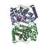


 PDBj
PDBj


 Assembly
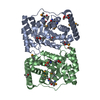

 Mass: 59.044 Da / Num. of mol.: 6 / Source method: obtained synthetically / Formula: C2H3O2
 Mass: 92.094 Da / Num. of mol.: 2 / Source method: obtained synthetically / Formula: C3H8O3
 Mass: 18.015 Da / Num. of mol.: 259 / Source method: isolated from a natural source / Formula: H2O
 Sample preparation
 / Beamline: BL9-2 / Wavelength: 0.89194,0.97946,0.97929
 Processing