 Movie
Movie Controller

[English] 日本語
 Yorodumi
- PDB-4e2w: X-ray Structure of the H181N mutant of TcaB9, a C-3'-Methyltransf... -
+ Open data

- Basic information
| Entry | Database: PDB / ID: 4e2w | ||||||
|---|---|---|---|---|---|---|---|
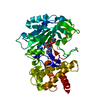
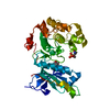
| Title | X-ray Structure of the H181N mutant of TcaB9, a C-3'-Methyltransferase, in Complex with S-Adenosyl-L-Homocysteine and Sugar Product | ||||||
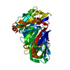
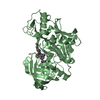
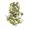
 Components Components | TcaB9 | ||||||
 Keywords Keywords | TRANSFERASE / kijanose / tetronitrose / tetradeoxy sugar / keto sugar / sugar methylation | ||||||
| Function / homology |  Function and homology information Function and homology information | ||||||
| Biological species |  Micromonospora chalcea (bacteria) Micromonospora chalcea (bacteria) | ||||||
| Method |  X-RAY DIFFRACTION / MOLECULAR REPLACEMENT / Resolution: 1.5 Å X-RAY DIFFRACTION / MOLECULAR REPLACEMENT / Resolution: 1.5 Å | ||||||
 Authors Authors | Bruender, N.A. / Holden, H.M. | ||||||
 Citation Citation | Journal: Protein Sci. / Year: 2012 Title: Probing the catalytic mechanism of a C-3'-methyltransferase involved in the biosynthesis of D-tetronitrose. Authors: Bruender, N.A. / Holden, H.M. | ||||||
| History |
|
- Structure visualization
| Structure viewer | Molecule: MolmilJmol/JSmol |
|---|
- Downloads & links
-Download
| PDBx/mmCIF format | 4e2w.cif.gz | 110.3 KB | Display | PDBx/mmCIF format |
|---|---|---|---|---|
| PDB format | pdb4e2w.ent.gz | 80.7 KB | Display | PDB format |
| PDBx/mmJSON format | 4e2w.json.gz | Tree view | PDBx/mmJSON format | |
| Others |  Other downloads Other downloads |
-Validation report
| Arichive directory | https://data.pdbj.org/pub/pdb/validation_reports/e2/4e2wftp://data.pdbj.org/pub/pdb/validation_reports/e2/4e2w | HTTPS FTP |
|---|
-Related structure data
| Related structure data |  4e2xC  4e2yC  4e2zC  4e30C 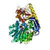 4e31C  4e32C 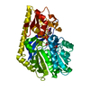 4e33C  3ndjS C: citing same article ( S: Starting model for refinement |
|---|---|
| Similar structure data |


-Links
 PDBj
PDBj- Assembly
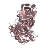
| Deposited unit | 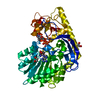
| ||||||||
|---|---|---|---|---|---|---|---|---|---|
| 1 |
| ||||||||
| Unit cell |
| ||||||||
| Components on special symmetry positions |
|
-Components
-Protein , 1 types, 1 molecules A
| #1: Protein | Mass: 46066.867 Da / Num. of mol.: 1 / Mutation: H181N Source method: isolated from a genetically manipulated source Source: (gene. exp.) Micromonospora chalcea (bacteria) / Gene: tcab9 / Plasmid: pET28 / Production host: References: UniProt: B5L6K6, Transferases; Transferring one-carbon groups; Methyltransferases |
|---|
-Non-polymers , 5 types, 447 molecules 


| #2: Chemical | ChemComp-ZN /  Mass: 65.409 Da / Num. of mol.: 1 / Source method: obtained synthetically / Formula: Zn Mass: 65.409 Da / Num. of mol.: 1 / Source method: obtained synthetically / Formula: Zn | ||||||
|---|---|---|---|---|---|---|---|
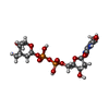
| #3: Chemical |  Mass: 62.068 Da / Num. of mol.: 3 / Source method: obtained synthetically / Formula: C2H6O2 Mass: 62.068 Da / Num. of mol.: 3 / Source method: obtained synthetically / Formula: C2H6O2#4: Chemical | ChemComp-SAH / |  Type: L-peptide linking / Mass: 384.411 Da / Num. of mol.: 1 / Source method: obtained synthetically / Formula: C14H20N6O5S Type: L-peptide linking / Mass: 384.411 Da / Num. of mol.: 1 / Source method: obtained synthetically / Formula: C14H20N6O5S#5: Chemical | ChemComp-JHZ / ( |  Mass: 543.356 Da / Num. of mol.: 1 / Source method: obtained synthetically / Formula: C17H27N3O13P2 Mass: 543.356 Da / Num. of mol.: 1 / Source method: obtained synthetically / Formula: C17H27N3O13P2#6: Water | ChemComp-HOH / | Mass: 18.015 Da / Num. of mol.: 441 / Source method: isolated from a natural source / Formula: H2O |
-Experimental details
-Experiment
| Experiment | Method: X-RAY DIFFRACTION / Number of used crystals: 1 |
|---|
- Sample preparation
| Crystal | Density Matthews: 2.37 Å3/Da / Density % sol: 48.19 % |
|---|---|
| Crystal grow | Temperature: 298 K / Method: vapor diffusion, hanging drop / pH: 8 Details: 1.2-1.6 M sodium/potassium phosphate, 10 mM dTMP, 5 mM S-adenosyl-L-homocysteine, pH 8.0, VAPOR DIFFUSION, HANGING DROP, temperature 298K |
-Data collection
| Diffraction | Mean temperature: 100 K |
|---|---|
| Diffraction source | Source: ROTATING ANODE / Type: RIGAKU RU200 / Wavelength: 1.54178 Å |
| Detector | Type: Bruker Platinum 135 / Detector: CCD / Date: Mar 25, 2011 / Details: Montel |
| Radiation | Monochromator: nickel filter / Protocol: SINGLE WAVELENGTH / Monochromatic (M) / Laue (L): M / Scattering type: x-ray |
| Radiation wavelength | Wavelength: 1.54178 Å / Relative weight: 1 |
| Reflection | Resolution: 1.5→38 Å / Num. all: 71732 / Num. obs: 70194 / % possible obs: 97.9 % / Redundancy: 4.7 % / Rmerge(I) obs: 0.084 / Rsym value: 0.084 / Net I/σ(I): 11.2 |
| Reflection shell | Resolution: 1.5→1.6 Å / Redundancy: 1.96 % / Rmerge(I) obs: 0.291 / Mean I/σ(I) obs: 2.77 / Num. unique all: 11310 / Rsym value: 0.291 / % possible all: 90.6 |
- Processing
| Software |
| |||||||||||||||||||||||||||||||||||||||||||||||||||||||||||||||||
|---|---|---|---|---|---|---|---|---|---|---|---|---|---|---|---|---|---|---|---|---|---|---|---|---|---|---|---|---|---|---|---|---|---|---|---|---|---|---|---|---|---|---|---|---|---|---|---|---|---|---|---|---|---|---|---|---|---|---|---|---|---|---|---|---|---|---|
| Refinement | Method to determine structure: MOLECULAR REPLACEMENT Starting model: PDB ENTRY 3NDJ Resolution: 1.5→38 Å / Cor.coef. Fo:Fc: 0.951 / Cor.coef. Fo:Fc free: 0.929 / SU B: 1.638 / SU ML: 0.059 / Cross valid method: THROUGHOUT / ESU R: 0.078 / ESU R Free: 0.083 / Stereochemistry target values: MAXIMUM LIKELIHOOD Details: HYDROGENS HAVE BEEN ADDED IN THE RIDING POSITIONS U VALUES : REFINED INDIVIDUALLY
| |||||||||||||||||||||||||||||||||||||||||||||||||||||||||||||||||
| Solvent computation | Ion probe radii: 0.8 Å / Shrinkage radii: 0.8 Å / VDW probe radii: 1.4 Å / Solvent model: MASK | |||||||||||||||||||||||||||||||||||||||||||||||||||||||||||||||||
| Displacement parameters | Biso mean: 12.769 Å2
| |||||||||||||||||||||||||||||||||||||||||||||||||||||||||||||||||
| Refinement step | Cycle: LAST / Resolution: 1.5→38 Å
| |||||||||||||||||||||||||||||||||||||||||||||||||||||||||||||||||
| Refine LS restraints |
| |||||||||||||||||||||||||||||||||||||||||||||||||||||||||||||||||
| LS refinement shell | Resolution: 1.5→1.537 Å / Total num. of bins used: 20
|
