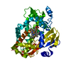
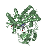
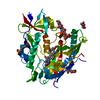
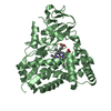
- PDB-4e2p: Crystal Structure of a Post-tailoring Hydroxylase (HmtN) Involved... -
+
Open data
ID or keywords:
Loading...
-
Basic information
Entry
Database: PDB / ID: 4e2p
Title
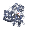
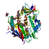
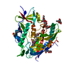
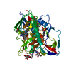
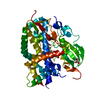
Crystal Structure of a Post-tailoring Hydroxylase (HmtN) Involved in the Himastatin Biosynthesis
Components
Cytochrome P450 107B1 (P450CVIIB1)
Keywords
OXIDOREDUCTASE / P450 / Hydroxylase / Oxygenase
Function / homology
Function and homology information
oxidoreductase activity, acting on paired donors, with incorporation or reduction of molecular oxygen / monooxygenase activity / iron ion binding / heme binding Similarity search - Function
Mass: 47109.574 Da / Num. of mol.: 1 Source method: isolated from a genetically manipulated source Source: (gene. exp.) Streptomyces himastatinicus (bacteria) / Strain: ATCC 53653 / Gene: hmtN / Plasmid: pET-28a / Production host: Escherichia coli (E. coli) / Strain (production host): BL21(DE3) References: UniProt: D9WMQ6, Oxidoreductases; Acting on single donors with incorporation of molecular oxygen (oxygenases); With incorporation of one atom of oxygen (internal monooxygenases or ...References: UniProt: D9WMQ6, Oxidoreductases; Acting on single donors with incorporation of molecular oxygen (oxygenases); With incorporation of one atom of oxygen (internal monooxygenases or internal mixed-function oxidases)
Resolution: 2.36→18.38 Å / Cor.coef. Fo:Fc: 0.89 / Cor.coef. Fo:Fc free: 0.854 / SU B: 17.032 / SU ML: 0.226 / Cross valid method: THROUGHOUT / σ(F): 0 / ESU R: 0.549 / ESU R Free: 0.301 / Stereochemistry target values: MAXIMUM LIKELIHOOD / Details: HYDROGENS HAVE BEEN USED IF PRESENT IN THE INPUT
Rfactor
Num. reflection
% reflection
Selection details
Rfree
0.28012
888
5.1 %
RANDOM
Rwork
0.23912
-
-
-
all
0.24121
17896
-
-
obs
0.24121
16424
96.48 %
-
Solvent computation
Ion probe radii: 0.8 Å / Shrinkage radii: 0.8 Å / VDW probe radii: 1.2 Å / Solvent model: MASK
Displacement parameters
Biso mean: 18.362 Å2
Baniso -1
Baniso -2
Baniso -3
1-
0.88 Å2
0 Å2
0 Å2
2-
-
-0.49 Å2
0 Å2
3-
-
-
-0.38 Å2
Refinement step
Cycle: LAST / Resolution: 2.36→18.38 Å
Protein
Nucleic acid
Ligand
Solvent
Total
Num. atoms
2939
0
47
219
3205
Refine LS restraints
Refine-ID
Type
Dev ideal
Dev ideal target
Number
X-RAY DIFFRACTION
r_bond_refined_d
0.005
0.019
3060
X-RAY DIFFRACTION
r_angle_refined_deg
1.458
1.991
4166
X-RAY DIFFRACTION
r_dihedral_angle_1_deg
3.839
5
367
X-RAY DIFFRACTION
r_dihedral_angle_2_deg
28.458
22.183
142
X-RAY DIFFRACTION
r_dihedral_angle_3_deg
12.6
15
499
X-RAY DIFFRACTION
r_dihedral_angle_4_deg
11.736
15
34
X-RAY DIFFRACTION
r_chiral_restr
0.052
0.2
449
X-RAY DIFFRACTION
r_gen_planes_refined
0.004
0.021
2361
LS refinement shell
Resolution: 2.358→2.418 Å / Total num. of bins used: 20
Rfactor
Num. reflection
% reflection
Rfree
0.308
53
-
Rwork
0.284
1054
-
obs
-
-
91.71 %
Refinement TLS params.
Method: refined / Origin x: 33.821 Å / Origin y: 67.1241 Å / Origin z: 17.3409 Å
11
12
13
21
22
23
31
32
33
T
0.0255 Å2
-0.0181 Å2
0.0041 Å2
-
0.0164 Å2
0.0029 Å2
-
-
0.0231 Å2
L
0.1426 °2
-0.2657 °2
0.0455 °2
-
0.617 °2
0.1185 °2
-
-
0.4268 °2
S
-0.0315 Å °
0.0106 Å °
0.0095 Å °
0.0273 Å °
0.0099 Å °
-0.0067 Å °
-0.0236 Å °
0.0346 Å °
0.0216 Å °
+
About Yorodumi
-
News
-
Feb 9, 2022. New format data for meta-information of EMDB entries
New format data for meta-information of EMDB entries
Version 3 of the EMDB header file is now the official format.
The previous official version 1.9 will be removed from the archive.
In the structure databanks used in Yorodumi, some data are registered as the other names, "COVID-19 virus" and "2019-nCoV". Here are the details of the virus and the list of structure data.
Jan 31, 2019. EMDB accession codes are about to change! (news from PDBe EMDB page)
EMDB accession codes are about to change! (news from PDBe EMDB page)
The allocation of 4 digits for EMDB accession codes will soon come to an end. Whilst these codes will remain in use, new EMDB accession codes will include an additional digit and will expand incrementally as the available range of codes is exhausted. The current 4-digit format prefixed with “EMD-” (i.e. EMD-XXXX) will advance to a 5-digit format (i.e. EMD-XXXXX), and so on. It is currently estimated that the 4-digit codes will be depleted around Spring 2019, at which point the 5-digit format will come into force.
The EM Navigator/Yorodumi systems omit the EMD- prefix.
Related info.:Q: What is EMD? / ID/Accession-code notation in Yorodumi/EM Navigator
Yorodumi is a browser for structure data from EMDB, PDB, SASBDB, etc.
This page is also the successor to EM Navigator detail page, and also detail information page/front-end page for Omokage search.
The word "yorodu" (or yorozu) is an old Japanese word meaning "ten thousand". "mi" (miru) is to see.
Related info.:EMDB / PDB / SASBDB / Comparison of 3 databanks / Yorodumi Search / Aug 31, 2016. New EM Navigator & Yorodumi / Yorodumi Papers / Jmol/JSmol / Function and homology information / Changes in new EM Navigator and Yorodumi
 Movie
Movie Controller

 Yorodumi
 Open data
 Basic information
 Components
 Keywords
 Function and homology information
 Streptomyces himastatinicus (bacteria)
 X-RAY DIFFRACTION /
  Authors
 Citation
 Structure visualization
 Downloads & links
 Other downloads
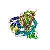

 PDBj
PDBj
 Assembly
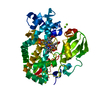

 Mass: 616.487 Da / Num. of mol.: 1 / Source method: obtained synthetically / Formula: C34H32FeN4O4
 Mass: 24.305 Da / Num. of mol.: 4 / Source method: obtained synthetically / Formula: Mg
 Mass: 18.015 Da / Num. of mol.: 219 / Source method: isolated from a natural source / Formula: H2O
 Sample preparation
 Processing