Resolution: 2.15→2.23 Å / Redundancy: 6.8 % / Mean I/σ(I) obs: 5.5 / Rsym value: 0.3 / % possible all: 98.3
-
Processing
Software
Name
Version
Classification
StructureStudio
datacollection
PHASER
phasing
REFMAC
5.6.0117
refinement
HKL-2000
datareduction
HKL-2000
datascaling
Refinement
Method to determine structure: MOLECULAR REPLACEMENT / Resolution: 2.15→41.94 Å / Cor.coef. Fo:Fc: 0.951 / Cor.coef. Fo:Fc free: 0.92 / SU B: 11.92 / SU ML: 0.155 / Cross valid method: THROUGHOUT / ESU R: 0.279 / ESU R Free: 0.215 / Stereochemistry target values: MAXIMUM LIKELIHOOD / Details: HYDROGENS HAVE BEEN USED IF PRESENT IN THE INPUT
Rfactor
Num. reflection
% reflection
Selection details
Rfree
0.24494
1686
5.1 %
RANDOM
Rwork
0.19003
-
-
-
all
0.19284
31670
-
-
obs
0.19284
31670
99.87 %
-
Solvent computation
Ion probe radii: 0.8 Å / Shrinkage radii: 0.8 Å / VDW probe radii: 1.2 Å / Solvent model: MASK
Displacement parameters
Biso mean: 30.924 Å2
Baniso -1
Baniso -2
Baniso -3
1-
0.23 Å2
0 Å2
-0.05 Å2
2-
-
-0.24 Å2
0 Å2
3-
-
-
-0.01 Å2
Refinement step
Cycle: LAST / Resolution: 2.15→41.94 Å
Protein
Nucleic acid
Ligand
Solvent
Total
Num. atoms
4811
0
40
197
5048
Refine LS restraints
Refine-ID
Type
Dev ideal
Dev ideal target
Number
X-RAY DIFFRACTION
r_bond_refined_d
0.017
0.02
5009
X-RAY DIFFRACTION
r_bond_other_d
X-RAY DIFFRACTION
r_angle_refined_deg
1.855
1.969
6792
X-RAY DIFFRACTION
r_angle_other_deg
X-RAY DIFFRACTION
r_dihedral_angle_1_deg
6.387
5
609
X-RAY DIFFRACTION
r_dihedral_angle_2_deg
34.896
25.041
244
X-RAY DIFFRACTION
r_dihedral_angle_3_deg
15.856
15
866
X-RAY DIFFRACTION
r_dihedral_angle_4_deg
18.315
15
16
X-RAY DIFFRACTION
r_chiral_restr
0.122
0.2
735
X-RAY DIFFRACTION
r_gen_planes_refined
0.009
0.021
3826
X-RAY DIFFRACTION
r_gen_planes_other
X-RAY DIFFRACTION
r_nbd_refined
X-RAY DIFFRACTION
r_nbd_other
X-RAY DIFFRACTION
r_nbtor_refined
X-RAY DIFFRACTION
r_nbtor_other
X-RAY DIFFRACTION
r_xyhbond_nbd_refined
X-RAY DIFFRACTION
r_xyhbond_nbd_other
X-RAY DIFFRACTION
r_metal_ion_refined
X-RAY DIFFRACTION
r_metal_ion_other
X-RAY DIFFRACTION
r_symmetry_vdw_refined
X-RAY DIFFRACTION
r_symmetry_vdw_other
X-RAY DIFFRACTION
r_symmetry_hbond_refined
X-RAY DIFFRACTION
r_symmetry_hbond_other
X-RAY DIFFRACTION
r_symmetry_metal_ion_refined
X-RAY DIFFRACTION
r_symmetry_metal_ion_other
X-RAY DIFFRACTION
r_mcbond_it
X-RAY DIFFRACTION
r_mcbond_other
X-RAY DIFFRACTION
r_mcangle_it
X-RAY DIFFRACTION
r_scbond_it
X-RAY DIFFRACTION
r_scangle_it
X-RAY DIFFRACTION
r_rigid_bond_restr
X-RAY DIFFRACTION
r_sphericity_free
X-RAY DIFFRACTION
r_sphericity_bonded
LS refinement shell
Resolution: 2.15→2.208 Å / Total num. of bins used: 20
Rfactor
Num. reflection
% reflection
Rfree
0.324
122
-
Rwork
0.233
2252
-
obs
-
-
98.34 %
Refinement TLS params.
Method: refined / Refine-ID: X-RAY DIFFRACTION
ID
L11 (°2)
L12 (°2)
L13 (°2)
L22 (°2)
L23 (°2)
L33 (°2)
S11 (Å °)
S12 (Å °)
S13 (Å °)
S21 (Å °)
S22 (Å °)
S23 (Å °)
S31 (Å °)
S32 (Å °)
S33 (Å °)
T11 (Å2)
T12 (Å2)
T13 (Å2)
T22 (Å2)
T23 (Å2)
T33 (Å2)
Origin x (Å)
Origin y (Å)
Origin z (Å)
1
3.5043
1.111
1.1515
0.6319
-0.5716
4.154
-0.0363
0.1328
-0.1419
-0.0541
-0.062
-0.1708
0.0434
0.344
0.0983
0.1248
-0.0216
0.0377
0.1529
0.0304
0.2008
16.3936
-5.5366
20.7039
2
1.6183
-0.0154
-0.3062
1.0122
0.1774
0.3246
0.0197
-0.0597
-0.0828
-0.0169
0.0421
0.0091
0.0359
-0.0576
-0.0618
0.079
-0.0319
-0.0298
0.1124
0.0206
0.0734
4.781
-10.1497
24.9932
3
1.3248
0.1938
0.4118
2.3276
1.3619
3.4264
0.0065
0.0232
0.0245
0.0933
-0.0766
0.1898
-0.1161
0.0748
0.0701
0.028
-0.0068
0.0044
0.0495
0.0157
0.0682
-9.9841
-9.1005
37.641
4
3.8339
-1.9139
1.059
2.3425
-0.5231
0.8632
0.0047
0.0628
-0.0997
-0.0368
-0.0143
0.1056
-0.0871
0.0933
0.0096
0.0528
0.0208
-0.02
0.0478
-0.0076
0.0912
-4.323
-39.2274
38.1879
5
1.6017
0.3525
-0.1044
4.4841
0.5754
1.4736
-0.0188
-0.0735
-0.0206
0.1021
0.0489
-0.0613
-0.0303
0.2434
-0.03
0.0141
0.0073
-0.0118
0.093
0.0174
0.0513
-6.2936
-10.6147
35.911
6
2.58
1.1264
0.929
4.5696
-0.9075
1.9794
0.2094
-0.3827
-0.1934
0.6265
-0.3348
0.1614
0.1073
-0.1081
0.1254
0.1465
-0.0284
0.0423
0.1317
0.0057
0.1139
-12.0144
-15.0953
44.9441
7
6.2343
-4.1761
-1.2016
5.5596
0.1786
0.3782
-0.2531
-0.2747
-0.0567
0.2376
0.2076
-0.1141
0.0173
0.0316
0.0454
0.1846
-0.0079
-0.0082
0.1287
-0.0304
0.0323
-0.9057
-29.7468
43.0279
8
10.4833
-2.2533
0.6524
1.0196
-0.6688
6.3289
-0.1163
-0.6109
0.4706
0.2915
0.0745
-0.8596
0.7152
0.9226
0.0418
0.4339
0.176
-0.4311
0.3508
-0.0784
1.2422
18.6982
-44.2322
23.8974
9
1.0412
0.4071
0.5059
2.7386
0.9564
1.9057
-0.0297
0.0028
-0.0565
-0.0832
0.0026
-0.3918
0.2204
0.0718
0.0271
0.0964
0.0048
0.0497
0.0485
0.0051
0.0786
8.7439
-40.9432
15.6026
10
1.3079
-0.3137
-0.0702
1.9608
1.4184
2.7864
0.0636
0.0724
-0.0993
0.0227
-0.0649
0.0557
-0.0553
-0.0219
0.0013
0.1092
-0.0369
0.0338
0.0358
-0.0041
0.0389
-0.9425
-43.7163
-1.4482
11
3.0216
0.4811
-0.3661
2.6489
-0.1714
0.7989
-0.0334
0.049
0.0447
0.0684
0.0506
0.3871
0.2399
0.2371
-0.0172
0.2121
0.0657
0.0218
0.083
0.0104
0.0956
0.8529
-12.7008
-0.3548
12
1.8076
-0.975
0.8073
4.5139
0.3699
1.6991
-0.0061
0.0265
-0.0083
0.1747
0.0168
-0.1249
-0.106
0.0583
-0.0107
0.1233
-0.0296
0.0268
0.0563
0.0018
0.0372
1.6371
-41.1599
1.7696
13
1.8172
0.5993
-0.8034
3.2623
-0.1799
1.2995
0.0227
0.2527
0.0658
-0.0634
-0.0433
0.0446
-0.2213
-0.0716
0.0206
0.1948
-0.0231
-0.0231
0.0893
-0.0039
0.0361
-0.8898
-37.3126
-8.7219
14
5.0918
4.0402
2.439
5.9191
2.3022
1.7644
-0.1892
0.1797
0.0461
-0.1746
0.3089
-0.3086
0.0667
0.1319
-0.1197
0.2614
0.0775
0.0594
0.1288
-0.0201
0.1284
6.8205
-22.4498
-2.9833
Refinement TLS group
ID
Refine-ID
Refine TLS-ID
Auth asym-ID
Auth seq-ID
1
X-RAY DIFFRACTION
1
A
1 - 23
2
X-RAY DIFFRACTION
2
A
24 - 97
3
X-RAY DIFFRACTION
3
A
98 - 144
4
X-RAY DIFFRACTION
4
A
145 - 189
5
X-RAY DIFFRACTION
5
A
190 - 225
6
X-RAY DIFFRACTION
6
A
226 - 253
7
X-RAY DIFFRACTION
7
A
254 - 301
8
X-RAY DIFFRACTION
8
B
1 - 23
9
X-RAY DIFFRACTION
9
B
24 - 97
10
X-RAY DIFFRACTION
10
B
98 - 144
11
X-RAY DIFFRACTION
11
B
145 - 189
12
X-RAY DIFFRACTION
12
B
190 - 225
13
X-RAY DIFFRACTION
13
B
226 - 253
14
X-RAY DIFFRACTION
14
B
254 - 296
+
About Yorodumi
-
News
-
Feb 9, 2022. New format data for meta-information of EMDB entries
New format data for meta-information of EMDB entries
Version 3 of the EMDB header file is now the official format.
The previous official version 1.9 will be removed from the archive.
In the structure databanks used in Yorodumi, some data are registered as the other names, "COVID-19 virus" and "2019-nCoV". Here are the details of the virus and the list of structure data.
Jan 31, 2019. EMDB accession codes are about to change! (news from PDBe EMDB page)
EMDB accession codes are about to change! (news from PDBe EMDB page)
The allocation of 4 digits for EMDB accession codes will soon come to an end. Whilst these codes will remain in use, new EMDB accession codes will include an additional digit and will expand incrementally as the available range of codes is exhausted. The current 4-digit format prefixed with “EMD-” (i.e. EMD-XXXX) will advance to a 5-digit format (i.e. EMD-XXXXX), and so on. It is currently estimated that the 4-digit codes will be depleted around Spring 2019, at which point the 5-digit format will come into force.
The EM Navigator/Yorodumi systems omit the EMD- prefix.
Related info.:Q: What is EMD? / ID/Accession-code notation in Yorodumi/EM Navigator
Yorodumi is a browser for structure data from EMDB, PDB, SASBDB, etc.
This page is also the successor to EM Navigator detail page, and also detail information page/front-end page for Omokage search.
The word "yorodu" (or yorozu) is an old Japanese word meaning "ten thousand". "mi" (miru) is to see.
Related info.:EMDB / PDB / SASBDB / Comparison of 3 databanks / Yorodumi Search / Aug 31, 2016. New EM Navigator & Yorodumi / Yorodumi Papers / Jmol/JSmol / Function and homology information / Changes in new EM Navigator and Yorodumi
 Movie
Movie Controller

 Yorodumi
 Open data
 Basic information
 Components
 Keywords
 Function and homology information
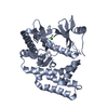
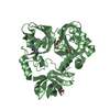
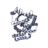
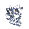
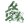
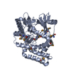
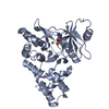
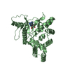
 Enterococcus casseliflavus (bacteria)
 X-RAY DIFFRACTION /
  Authors
 Citation
 Structure visualization
 Downloads & links
 Other downloads


 PDBj
PDBj

 Assembly
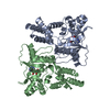

 Mass: 267.241 Da / Num. of mol.: 2 / Source method: obtained synthetically / Formula: C10H13N5O4
 Mass: 35.453 Da / Num. of mol.: 2 / Source method: obtained synthetically / Formula: Cl
 Mass: 18.015 Da / Num. of mol.: 197 / Source method: isolated from a natural source / Formula: H2O
 Sample preparation
 Processing