 Movie
Movie Controller

[English] 日本語
 Yorodumi
- PDB-4dt0: The structure of the peripheral stalk subunit E from Pyrococcus h... -
+ Open data

- Basic information
| Entry | Database: PDB / ID: 4dt0 | ||||||
|---|---|---|---|---|---|---|---|
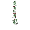
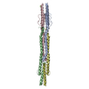
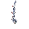
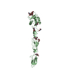
| Title | The structure of the peripheral stalk subunit E from Pyrococcus horikoshii | ||||||
 Components Components | V-type ATP synthase subunit E | ||||||
 Keywords Keywords | HYDROLASE / A-ATP synthase / peripheral stalk | ||||||
| Function / homology |  Function and homology information Function and homology informationproton-transporting two-sector ATPase complex, catalytic domain / proton motive force-driven plasma membrane ATP synthesis / proton-transporting ATPase activity, rotational mechanism / proton-transporting ATP synthase activity, rotational mechanism / ATP binding / plasma membrane Similarity search - Function | ||||||
| Biological species |   Pyrococcus horikoshii (archaea) Pyrococcus horikoshii (archaea) | ||||||
| Method |  X-RAY DIFFRACTION / SYNCHROTRON / MAD / Resolution: 3.65 Å X-RAY DIFFRACTION / SYNCHROTRON / MAD / Resolution: 3.65 Å | ||||||
 Authors Authors | Balakrishna, A.M. / Gruber, G. | ||||||
 Citation Citation | Journal: J.Mol.Biol. / Year: 2012 Title: The structure of subunit E of the Pyrococcus horikoshii OT3 A-ATP synthase gives insight into the elasticity of the peripheral stalk. Authors: Balakrishna, A.M. / Hunke, C. / Gruber, G. #1: Journal: J.Mol.Biol. / Year: 2007 Title: Dimeric core structure of modular stator subunit E of archaeal H+ -ATPase. Authors: Lokanath, N.K. / Matsuura, Y. / Kuroishi, C. / Takahashi, N. / Kunishima, N. | ||||||
| History |
|
- Structure visualization
| Structure viewer | Molecule: MolmilJmol/JSmol |
|---|
- Downloads & links
-Download
| PDBx/mmCIF format | 4dt0.cif.gz | 55.6 KB | Display | PDBx/mmCIF format |
|---|---|---|---|---|
| PDB format | pdb4dt0.ent.gz | 37.2 KB | Display | PDB format |
| PDBx/mmJSON format | 4dt0.json.gz | Tree view | PDBx/mmJSON format | |
| Others |  Other downloads Other downloads |
-Validation report
| Arichive directory | https://data.pdbj.org/pub/pdb/validation_reports/dt/4dt0ftp://data.pdbj.org/pub/pdb/validation_reports/dt/4dt0 | HTTPS FTP |
|---|
-Related structure data
| Related structure data | |
|---|---|
| Similar structure data |


-Links
 PDBj
PDBj

- Assembly
| Deposited unit | 
| ||||||||
|---|---|---|---|---|---|---|---|---|---|
| 1 |
| ||||||||
| Unit cell |
|
-Components
| #1: Protein | Mass: 24182.723 Da / Num. of mol.: 1 Source method: isolated from a genetically manipulated source Source: (gene. exp.) Pyrococcus horikoshii (archaea) / Strain: OT3 / Gene: atpE, PH1978 / Plasmid: pET22b(+)-His6 / Production host:  |
|---|
-Experimental details
-Experiment
| Experiment | Method: X-RAY DIFFRACTION / Number of used crystals: 1 |
|---|
- Sample preparation
| Crystal | Density Matthews: 3.25 Å3/Da / Density % sol: 62.21 % |
|---|---|
| Crystal grow | Temperature: 298 K / Method: vapor diffusion, sitting drop / pH: 6 Details: 10% 2-propanol, 100mM 2-morpholinoethanesulfonic acid, sodium salt (pH 6.0), 200mM calcium acetate, 0.01 M potassium sodium tartrate tetrahydrate, VAPOR DIFFUSION, SITTING DROP, temperature 298K |
-Data collection
| Diffraction | Mean temperature: 100 K | ||||||||||||
|---|---|---|---|---|---|---|---|---|---|---|---|---|---|
| Diffraction source | Source: SYNCHROTRON / Site: NSRRC  / Beamline: BL13B1 / Wavelength: 0.979, 0.978, 0.963 / Beamline: BL13B1 / Wavelength: 0.979, 0.978, 0.963 | ||||||||||||
| Detector | Type: ADSC QUANTUM 315 / Detector: CCD / Date: Jun 24, 2011 Details: Vertically Collimating Premirror, Toroidal Focusing Mirror | ||||||||||||
| Radiation | Monochromator: Double Crystal Si(111) Monochromator / Protocol: MAD / Monochromatic (M) / Laue (L): M / Scattering type: x-ray | ||||||||||||
| Radiation wavelength |
| ||||||||||||
| Reflection | Resolution: 3.65→30 Å / Num. all: 3599 / Num. obs: 3564 / % possible obs: 99 % / Observed criterion σ(F): 0 / Observed criterion σ(I): 0 / Redundancy: 4.1 % / Biso Wilson estimate: 88.63 Å2 / Rmerge(I) obs: 0.06 / Net I/σ(I): 13.5 | ||||||||||||
| Reflection shell | Resolution: 3.65→3.78 Å / Redundancy: 4 % / Rmerge(I) obs: 0.38 / Mean I/σ(I) obs: 2.08 / Num. unique all: 3599 / % possible all: 99.7 |
- Processing
| Software |
| ||||||||||||||||||||||||||||||||||||||||
|---|---|---|---|---|---|---|---|---|---|---|---|---|---|---|---|---|---|---|---|---|---|---|---|---|---|---|---|---|---|---|---|---|---|---|---|---|---|---|---|---|---|
| Refinement | Method to determine structure: MAD / Resolution: 3.65→26.85 Å / Cor.coef. Fo:Fc: 0.887 / Cor.coef. Fo:Fc free: 0.861 / SU B: 128.307 / SU ML: 0.898 / Cross valid method: THROUGHOUT / ESU R Free: 0.748 / Stereochemistry target values: MAXIMUM LIKELIHOOD / Details: HYDROGENS HAVE BEEN ADDED IN THE RIDING POSITIONS
| ||||||||||||||||||||||||||||||||||||||||
| Solvent computation | Ion probe radii: 0.8 Å / Shrinkage radii: 0.8 Å / VDW probe radii: 1.4 Å / Solvent model: MASK | ||||||||||||||||||||||||||||||||||||||||
| Displacement parameters | Biso mean: 72.928 Å2
| ||||||||||||||||||||||||||||||||||||||||
| Refinement step | Cycle: LAST / Resolution: 3.65→26.85 Å
| ||||||||||||||||||||||||||||||||||||||||
| Refine LS restraints |
| ||||||||||||||||||||||||||||||||||||||||
| LS refinement shell | Resolution: 3.651→3.744 Å / Total num. of bins used: 20
| ||||||||||||||||||||||||||||||||||||||||
| Refinement TLS params. | Method: refined / Origin x: 11.0024 Å / Origin y: -9.924 Å / Origin z: 0.3318 Å
|
