 Movie
Movie Controller

[English] 日本語
 Yorodumi
- PDB-4csy: E-selectin lectin, EGF-like and two SCR domains complexed with Si... -
+ Open data

- Basic information
| Entry | Database: PDB / ID: 4csy | |||||||||
|---|---|---|---|---|---|---|---|---|---|---|
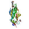
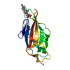
| Title | E-selectin lectin, EGF-like and two SCR domains complexed with Sialyl Lewis X | |||||||||
 Components Components | E-SELECTIN | |||||||||
 Keywords Keywords | CELL ADHESION / CELL-ADHESION / HUMAN LECTIN / C-TYPE LECTIN / INFLAMMATION / LEUKOCYTE / SIALYL LEWIS X / SLEX / PROTEIN CONFORMATION / LIGAND-INDUCED CONFORMATIONAL CHANGE / CATCH- BOND | |||||||||
| Function / homology |  Function and homology information Function and homology informationactin filament-based process / positive regulation of leukocyte tethering or rolling / sialic acid binding / leukocyte migration involved in inflammatory response / oligosaccharide binding / leukocyte tethering or rolling / positive regulation of leukocyte migration / heterophilic cell-cell adhesion / leukocyte cell-cell adhesion / cortical cytoskeleton ...actin filament-based process / positive regulation of leukocyte tethering or rolling / sialic acid binding / leukocyte migration involved in inflammatory response / oligosaccharide binding / leukocyte tethering or rolling / positive regulation of leukocyte migration / heterophilic cell-cell adhesion / leukocyte cell-cell adhesion / cortical cytoskeleton / positive regulation of receptor internalization / response to tumor necrosis factor / phospholipase binding / clathrin-coated pit / response to cytokine / response to interleukin-1 / Cell surface interactions at the vascular wall / calcium-mediated signaling / caveola / transmembrane signaling receptor activity / regulation of inflammatory response / response to lipopolysaccharide / phospholipase C-activating G protein-coupled receptor signaling pathway / membrane raft / inflammatory response / external side of plasma membrane / perinuclear region of cytoplasm / : / metal ion binding / plasma membrane Similarity search - Function | |||||||||
| Biological species |  HOMO SAPIENS (human) HOMO SAPIENS (human) | |||||||||
| Method |  X-RAY DIFFRACTION / SYNCHROTRON / MOLECULAR REPLACEMENT / Resolution: 2.41 Å X-RAY DIFFRACTION / SYNCHROTRON / MOLECULAR REPLACEMENT / Resolution: 2.41 Å | |||||||||
 Authors Authors | Preston, R.C. / Jakob, R.P. / Binder, F.P.C. / Sager, C.P. / Ernst, B. / Maier, T. | |||||||||
 Citation Citation | Journal: J.Mol.Cell.Biol. / Year: 2016 Title: E-Selectin Ligand Complexes Adopt an Extended High-Affinity Conformation. Authors: Preston, R.C. / Jakob, R.P. / Binder, F.P. / Sager, C.P. / Ernst, B. / Maier, T. | |||||||||
| History |
|
- Structure visualization
| Structure viewer | Molecule: MolmilJmol/JSmol |
|---|
- Downloads & links
-Download
| PDBx/mmCIF format | 4csy.cif.gz | 254.3 KB | Display | PDBx/mmCIF format |
|---|---|---|---|---|
| PDB format | pdb4csy.ent.gz | 207.3 KB | Display | PDB format |
| PDBx/mmJSON format | 4csy.json.gz | Tree view | PDBx/mmJSON format | |
| Others |  Other downloads Other downloads |
-Validation report
| Arichive directory | https://data.pdbj.org/pub/pdb/validation_reports/cs/4csyftp://data.pdbj.org/pub/pdb/validation_reports/cs/4csy | HTTPS FTP |
|---|
-Related structure data
| Related structure data |  4c16C 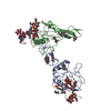 1g1sS 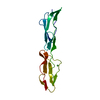 1h04S  3govS S: Starting model for refinement C: citing same article ( |
|---|---|
| Similar structure data |


-Links
 PDBj
PDBj


- Assembly
| Deposited unit | 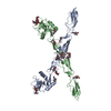
| ||||||||
|---|---|---|---|---|---|---|---|---|---|
| 1 | 
| ||||||||
| 2 | 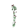
| ||||||||
| Unit cell |
| ||||||||
| Noncrystallographic symmetry (NCS) | NCS oper: (Code: given Matrix: (-1, -0.005513, -0.005256), Vector: |
-Components
| #1: Protein | Mass: 31305.760 Da / Num. of mol.: 2 Fragment: LECTIN DOMAIN, EGF-LIKE DOMAIN, SHORT CONSENSUS REPEAT DOMAIN 1, SHORT CONSENSUS REPEAT DOMAIN 2, RESIDUES 22-301 Source method: isolated from a genetically manipulated source Details: N-ACETYLGLUCOSAMINE RESIDUES ATTACHED TO ASN4, ASN124, ASN139, ASN158, ASN178, ASN182, AND ASN244 ON BOTH CHAINS. Source: (gene. exp.) HOMO SAPIENS (human) / Tissue: CYTOKINE-INDUCED VASCULAR ENDOTHELIAL CELLS / Plasmid: PCDNA3.1Cell line (production host): CHINESE HAMSTER OVARY (CHO) CELLS Production host:   CRICETULUS GRISEUS (Chinese hamster) / Tissue (production host): OVARY / References: UniProt: P16581 CRICETULUS GRISEUS (Chinese hamster) / Tissue (production host): OVARY / References: UniProt: P16581#2: Polysaccharide | Source method: isolated from a genetically manipulated source #3: Sugar | ChemComp-NAG /   Type: D-saccharide, beta linking / Mass: 221.208 Da / Num. of mol.: 14 Type: D-saccharide, beta linking / Mass: 221.208 Da / Num. of mol.: 14Source method: isolated from a genetically manipulated source Formula: C8H15NO6 #4: Chemical |   Mass: 40.078 Da / Num. of mol.: 2 / Source method: obtained synthetically / Formula: Ca Mass: 40.078 Da / Num. of mol.: 2 / Source method: obtained synthetically / Formula: Ca#5: Water | ChemComp-HOH / |  Mass: 18.015 Da / Num. of mol.: 127 / Source method: isolated from a natural source / Formula: H2O Mass: 18.015 Da / Num. of mol.: 127 / Source method: isolated from a natural source / Formula: H2OHas protein modification | Y | Nonpolymer details | SIALYL LEWIS X METHYL GLUCOSIDE (DRG): SIALYL-LEWIS-X TETRASACCHARIDE, WITH A TRIMETHYLSILYLETHYL ...SIALYL LEWIS X METHYL GLUCOSIDE (DRG): SIALYL-LEWIS-X TETRASACCH | Sequence details | WITHOUT N-TERMINAL SECRETION SIGNAL (AA. 1-21). SEQUENCE OF MATURE PROTEIN STARTS WITH RESIDUE 1 ...WITHOUT N-TERMINAL SECRETION SIGNAL (AA. 1-21). SEQUENCE OF MATURE PROTEIN STARTS WITH RESIDUE 1 FOR COMPATIBIL | |
|---|
-Experimental details
-Experiment
| Experiment | Method: X-RAY DIFFRACTION / Number of used crystals: 1 |
|---|
- Sample preparation
| Crystal | Density Matthews: 2.8 Å3/Da / Density % sol: 56.1 % / Description: NONE |
|---|---|
| Crystal grow | Details: 13 % PEG8000, HEPES, MOPS PH 6.2, CACL2, 10MM SLEX-OTMSE |
-Data collection
| Diffraction | Mean temperature: 100 K |
|---|---|
| Diffraction source | Source: SYNCHROTRON / Site: SLS  / Beamline: X06SA / Wavelength: 0.99985 / Beamline: X06SA / Wavelength: 0.99985 |
| Detector | Type: DECTRIS PILATUS 6M / Detector: PIXEL / Date: Mar 30, 2012 |
| Radiation | Protocol: SINGLE WAVELENGTH / Monochromatic (M) / Laue (L): M / Scattering type: x-ray |
| Radiation wavelength | Wavelength: 0.99985 Å / Relative weight: 1 |
| Reflection | Resolution: 2.41→28.28 Å / Num. obs: 25239 / % possible obs: 95.4 % / Observed criterion σ(I): 2 / Redundancy: 1.8 % / Biso Wilson estimate: 66.77 Å2 / Rmerge(I) obs: 0.04 / Net I/σ(I): 8.8 |
| Reflection shell | Resolution: 2.41→2.53 Å / Redundancy: 1.8 % / Rmerge(I) obs: 0.32 / Mean I/σ(I) obs: 2 / % possible all: 90.2 |
- Processing
| Software |
| ||||||||||||||||||||||||||||||||||||||||||||||||||||||||||||||||||||||||||||||||||||||||||||||||||||||||||||||||||
|---|---|---|---|---|---|---|---|---|---|---|---|---|---|---|---|---|---|---|---|---|---|---|---|---|---|---|---|---|---|---|---|---|---|---|---|---|---|---|---|---|---|---|---|---|---|---|---|---|---|---|---|---|---|---|---|---|---|---|---|---|---|---|---|---|---|---|---|---|---|---|---|---|---|---|---|---|---|---|---|---|---|---|---|---|---|---|---|---|---|---|---|---|---|---|---|---|---|---|---|---|---|---|---|---|---|---|---|---|---|---|---|---|---|---|---|
| Refinement | Method to determine structure: MOLECULAR REPLACEMENT Starting model: PDB ENTRIES 1G1S, 3GOV, 1H04 Resolution: 2.41→28.32 Å / Cor.coef. Fo:Fc: 0.9294 / Cor.coef. Fo:Fc free: 0.9003 / SU R Cruickshank DPI: 0.471 / Cross valid method: THROUGHOUT / σ(F): 0 / SU R Blow DPI: 0.442 / SU Rfree Blow DPI: 0.261 / SU Rfree Cruickshank DPI: 0.268 Details: DISORDER IN LOOPS 227-231, 254-259 AND N-TERMINAL RESIDUES 278-280 BUILT ACCORDING TO ACCOMPANYING DEPOSITION EBI-57874.
| ||||||||||||||||||||||||||||||||||||||||||||||||||||||||||||||||||||||||||||||||||||||||||||||||||||||||||||||||||
| Displacement parameters | Biso mean: 79.3 Å2
| ||||||||||||||||||||||||||||||||||||||||||||||||||||||||||||||||||||||||||||||||||||||||||||||||||||||||||||||||||
| Refine analyze | Luzzati coordinate error obs: 0.507 Å | ||||||||||||||||||||||||||||||||||||||||||||||||||||||||||||||||||||||||||||||||||||||||||||||||||||||||||||||||||
| Refinement step | Cycle: LAST / Resolution: 2.41→28.32 Å
| ||||||||||||||||||||||||||||||||||||||||||||||||||||||||||||||||||||||||||||||||||||||||||||||||||||||||||||||||||
| Refine LS restraints |
| ||||||||||||||||||||||||||||||||||||||||||||||||||||||||||||||||||||||||||||||||||||||||||||||||||||||||||||||||||
| LS refinement shell | Resolution: 2.41→2.51 Å / Total num. of bins used: 13
| ||||||||||||||||||||||||||||||||||||||||||||||||||||||||||||||||||||||||||||||||||||||||||||||||||||||||||||||||||
| Refinement TLS params. | Method: refined / Refine-ID: X-RAY DIFFRACTION
| ||||||||||||||||||||||||||||||||||||||||||||||||||||||||||||||||||||||||||||||||||||||||||||||||||||||||||||||||||
| Refinement TLS group |
|
