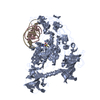
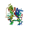
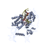
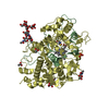
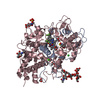
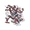
- PDB-4dkj: CpG specific methyltransferase in complex with target DNA -
+
Open data
ID or keywords:
Loading...
-
Basic information
Entry
Database: PDB / ID: 4dkj
Title
CpG specific methyltransferase in complex with target DNA
Components
Cytosine-specific methyltransferase
DNA (5'-D(*CP*CP*AP*CP*AP*TP*GP*(C37)P*GP*CP*TP*GP*AP*A)-3')
DNA (5'-D(*GP*TP*TP*CP*AP*GP*(5CM)P*GP*CP*AP*TP*GP*TP*G)-3')
Keywords
TRANSFERASE/DNA / CG-SPECIFICITY / DNA INTERCALATION / CPG SEQUENCE / CYTOSINE C5-METHYLATION / C5-METHYLCYTOSINE / NUCLEOTIDE FLIPPING / S-adenosyl-L-methionine-dependent methyltransferases / C-5 cytosine-specific DNA methylases / DNA (cytosine-5-)-methyltransferase activity / DNA binding / DNA (cytosine-5-)-methylation / intracellular / TRANSFERASE-DNA complex
Function / homology
Function and homology information
DNA (cytosine-5-)-methyltransferase / DNA (cytosine-5-)-methyltransferase activity / DNA restriction-modification system / methylation Similarity search - Function
: / DNA Methylase, subunit A, domain 2 / DNA Methylase; Chain A, domain 2 / DNA methylase, C-5 cytosine-specific, active site / C-5 cytosine-specific DNA methylases active site. / C-5 cytosine-specific DNA methylase (Dnmt) domain profile. / C-5 cytosine methyltransferase / C-5 cytosine-specific DNA methylase / Vaccinia Virus protein VP39 / S-adenosyl-L-methionine-dependent methyltransferase superfamily ...: / DNA Methylase, subunit A, domain 2 / DNA Methylase; Chain A, domain 2 / DNA methylase, C-5 cytosine-specific, active site / C-5 cytosine-specific DNA methylases active site. / C-5 cytosine-specific DNA methylase (Dnmt) domain profile. / C-5 cytosine methyltransferase / C-5 cytosine-specific DNA methylase / Vaccinia Virus protein VP39 / S-adenosyl-L-methionine-dependent methyltransferase superfamily / Alpha-Beta Complex / Rossmann fold / 3-Layer(aba) Sandwich / Alpha Beta Similarity search - Domain/homology
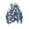
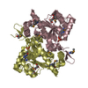
AUTHORS STATE THAT THE BIOLOGICAL UNIT CONTAINS PROTEIN CHAIN A, AND DNA STRANDS B AND C. THE TRIMER (ACCORDING TO PDB CONVENTIONS) IS A COMPLEX OF THE MONOMERIC METHYLTRANSFERASE WITH ITS SUBSTRATE, DOUBLE STRANDED DNA.
-
Components
-
Protein , 1 types, 1 molecules A
#1: Protein
Cytosine-specificmethyltransferase
Mass: 47310.082 Da / Num. of mol.: 1 / Mutation: S295P Source method: isolated from a genetically manipulated source Source: (gene. exp.) Mycoplasma penetrans (bacteria) / Strain: HF-2 / Gene: MYPE4940 / Plasmid: PET28A / Production host: Escherichia coli (E. coli) / Strain (production host): ER2566 References: UniProt: Q8EVR5, DNA (cytosine-5-)-methyltransferase
-
DNA chain , 2 types, 2 molecules BC
#2: DNA chain
DNA (5'-D(*CP*CP*AP*CP*AP*TP*GP*(C37)P*GP*CP*TP*GP*AP*A)-3')
Mass: 4267.773 Da / Num. of mol.: 1 / Source method: obtained synthetically
#3: DNA chain
DNA (5'-D(*GP*TP*TP*CP*AP*GP*(5CM)P*GP*CP*AP*TP*GP*TP*G)-3')
Mass: 4325.829 Da / Num. of mol.: 1 / Source method: obtained synthetically
Mass: 18.015 Da / Num. of mol.: 267 / Source method: isolated from a natural source / Formula: H2O
-
Details
Has protein modification
N
Sequence details
AUTHORS STATE THAT THESE ARE NATURAL STRAIN VARIATIONS.
-
Experimental details
-
Experiment
Experiment
Method: X-RAY DIFFRACTION / Number of used crystals: 1
-
Sample preparation
Crystal
Density Matthews: 2.74 Å3/Da / Density % sol: 55.09 %
Crystal grow
Temperature: 294 K / Method: vapor diffusion, sitting drop / pH: 5.6 Details: 10% PEG 3350, 150 mM NaCl, 50 mM sodium. For cryoprotection glycerol was added to 25% v/v, pH 5.6, VAPOR DIFFUSION, SITTING DROP, temperature 294K
Resolution: 2.15→38.6 Å / Cor.coef. Fo:Fc: 0.954 / Cor.coef. Fo:Fc free: 0.935 / Cross valid method: THROUGHOUT / ESU R: 0.214 / ESU R Free: 0.176 / Stereochemistry target values: MAXIMUM LIKELIHOOD Details: (1) THE CRYSTAL WAS GROWN IN THE PRESENCE OF A SAMPLE THAT WAS LABELLED AS S-ADENOSYL-METHIONINE (SAM), BUT APPEARS TO HAVE BEEN DEGRADED TO S-ADENOSYL-HOMOCYSTEINE (SAH) EITHER PRIOR TO OR ...Details: (1) THE CRYSTAL WAS GROWN IN THE PRESENCE OF A SAMPLE THAT WAS LABELLED AS S-ADENOSYL-METHIONINE (SAM), BUT APPEARS TO HAVE BEEN DEGRADED TO S-ADENOSYL-HOMOCYSTEINE (SAH) EITHER PRIOR TO OR DURING CRYSTALLIZATION. PROGRAM CNS HAS BEEN USED FOR DNA REFINEMENT. NO SUGAR PUCKER CONSTRAINTS HAVE BEEN APPLIED. (2) THE MODELLED S-ADENOSYLHOMOCYSTEINE (SAH) IS UNCERTAIN. ITS BINDING SITE MIGHT BE OCCUPIED IN PART BY S-ADENOSYLMETHIONINE (SAM) OR THE SAM DEGRADATION PRODUCT 5 -METHYLTHIOADENOSINE (MTA) (FROM SCISSION OF SAM TO MTA AND HOMOSERINE LACTONE). THE DISTANCE BETWEEN CYS135 S AND C6 OF THE SUBSTRATE CYTOSINE BASE IS INTERMEDIATE BETWEEN A NON-COVALENT AND A COVALENT BOND. A DISTANCE BELOW THE VAN DER WAALS LIMIT SUGGESTS THAT SLIGHT DEVIATIONS FROM THE PLANARITY OF THE DNA BASE SHOULD OCCUR. AS THE RESOLUTION OF THE X-RAY DATA IS INSUFFICIENT TO DETECT SUCH DEVIATIONS, THEY WERE NOT MODELLED. (3) HYDROGENS HAVE BEEN ADDED IN THE RIDING POSITIONS. TLS REFINEMENT HAS BEEN USED.
Rfactor
Num. reflection
% reflection
Selection details
Rfree
0.21631
1729
5 %
RANDOM
Rwork
0.17632
-
-
-
all
0.17832
34274
-
-
obs
0.17832
34274
98.96 %
-
Solvent computation
Ion probe radii: 0.8 Å / Shrinkage radii: 0.8 Å / VDW probe radii: 1.4 Å / Solvent model: MASK
Refinement step
Cycle: LAST / Resolution: 2.15→38.6 Å
Protein
Nucleic acid
Ligand
Solvent
Total
Num. atoms
3199
570
32
267
4068
Refine LS restraints
Refine-ID
Type
Dev ideal
Dev ideal target
Number
X-RAY DIFFRACTION
r_bond_refined_d
0.01
0.022
4388
X-RAY DIFFRACTION
r_bond_other_d
0
0.02
2941
X-RAY DIFFRACTION
r_angle_refined_deg
1.253
2.131
6064
X-RAY DIFFRACTION
r_angle_other_deg
3.966
3
7256
X-RAY DIFFRACTION
r_dihedral_angle_1_deg
6.16
5
471
X-RAY DIFFRACTION
r_dihedral_angle_2_deg
38.254
25.301
183
X-RAY DIFFRACTION
r_dihedral_angle_3_deg
15.063
15
767
X-RAY DIFFRACTION
r_dihedral_angle_4_deg
15.209
15
17
X-RAY DIFFRACTION
r_chiral_restr
X-RAY DIFFRACTION
r_gen_planes_refined
0.007
0.02
4548
X-RAY DIFFRACTION
r_gen_planes_other
0.007
0.02
810
X-RAY DIFFRACTION
r_nbd_refined
0.192
0.2
771
X-RAY DIFFRACTION
r_nbd_other
0.232
0.2
2985
X-RAY DIFFRACTION
r_nbtor_refined
0.181
0.2
2059
X-RAY DIFFRACTION
r_nbtor_other
0.108
0.2
1868
X-RAY DIFFRACTION
r_xyhbond_nbd_refined
0.137
0.2
258
X-RAY DIFFRACTION
r_xyhbond_nbd_other
X-RAY DIFFRACTION
r_metal_ion_refined
X-RAY DIFFRACTION
r_metal_ion_other
X-RAY DIFFRACTION
r_symmetry_vdw_refined
0.079
0.2
14
X-RAY DIFFRACTION
r_symmetry_vdw_other
0.38
0.2
15
X-RAY DIFFRACTION
r_symmetry_hbond_refined
0.084
0.2
3
X-RAY DIFFRACTION
r_symmetry_hbond_other
X-RAY DIFFRACTION
r_symmetry_metal_ion_refined
X-RAY DIFFRACTION
r_symmetry_metal_ion_other
X-RAY DIFFRACTION
r_mcbond_it
X-RAY DIFFRACTION
r_mcbond_other
X-RAY DIFFRACTION
r_mcangle_it
X-RAY DIFFRACTION
r_scbond_it
X-RAY DIFFRACTION
r_scangle_it
X-RAY DIFFRACTION
r_rigid_bond_restr
X-RAY DIFFRACTION
r_sphericity_free
X-RAY DIFFRACTION
r_sphericity_bonded
LS refinement shell
Resolution: 2.15→2.206 Å / Total num. of bins used: 20
Rfactor
Num. reflection
% reflection
Rfree
0.288
138
-
Rwork
0.203
2348
-
obs
-
2486
99.92 %
Refinement TLS params.
Method: refined / Refine-ID: X-RAY DIFFRACTION
ID
L11 (°2)
L12 (°2)
L13 (°2)
L22 (°2)
L23 (°2)
L33 (°2)
S11 (Å °)
S12 (Å °)
S13 (Å °)
S21 (Å °)
S22 (Å °)
S23 (Å °)
S31 (Å °)
S32 (Å °)
S33 (Å °)
T11 (Å2)
T12 (Å2)
T13 (Å2)
T22 (Å2)
T23 (Å2)
T33 (Å2)
Origin x (Å)
Origin y (Å)
Origin z (Å)
1
1.3285
0.7139
0.0899
1.8304
0.216
1.446
0.1138
-0.2095
0.1597
0.0245
-0.0982
0.065
-0.1703
0.1231
-0.0156
-0.0916
-0.0668
0.0118
-0.0382
-0.023
-0.0857
25.875
30.823
49.244
2
0.9724
0.6691
-0.1999
2.2069
0.0468
0.7196
-0.0188
-0.0366
-0.0063
-0.1209
-0.0562
0.1295
0.0652
-0.0767
0.0749
-0.0695
-0.0421
-0.0339
-0.0185
0.0294
-0.026
12.601
9.256
40.543
3
1.3002
-0.0789
0.4393
1.9057
-0.0269
2.1045
0.1105
-0.0305
-0.0781
-0.0129
0.0146
-0.2426
0.1346
0.3025
-0.1251
-0.0503
0.0155
0.0014
0.0055
0.0614
-0.0227
29.06
5.76
43.562
Refinement TLS group
ID
Refine-ID
Refine TLS-ID
Auth asym-ID
Auth seq-ID
1
X-RAY DIFFRACTION
1
A
6 - 259
2
X-RAY DIFFRACTION
1
A
376 - 393
3
X-RAY DIFFRACTION
2
A
260 - 375
4
X-RAY DIFFRACTION
3
B
1 - 14
5
X-RAY DIFFRACTION
3
C
1 - 14
+
About Yorodumi
-
News
-
Feb 9, 2022. New format data for meta-information of EMDB entries
New format data for meta-information of EMDB entries
Version 3 of the EMDB header file is now the official format.
The previous official version 1.9 will be removed from the archive.
In the structure databanks used in Yorodumi, some data are registered as the other names, "COVID-19 virus" and "2019-nCoV". Here are the details of the virus and the list of structure data.
Jan 31, 2019. EMDB accession codes are about to change! (news from PDBe EMDB page)
EMDB accession codes are about to change! (news from PDBe EMDB page)
The allocation of 4 digits for EMDB accession codes will soon come to an end. Whilst these codes will remain in use, new EMDB accession codes will include an additional digit and will expand incrementally as the available range of codes is exhausted. The current 4-digit format prefixed with “EMD-” (i.e. EMD-XXXX) will advance to a 5-digit format (i.e. EMD-XXXXX), and so on. It is currently estimated that the 4-digit codes will be depleted around Spring 2019, at which point the 5-digit format will come into force.
The EM Navigator/Yorodumi systems omit the EMD- prefix.
Related info.:Q: What is EMD? / ID/Accession-code notation in Yorodumi/EM Navigator
Yorodumi is a browser for structure data from EMDB, PDB, SASBDB, etc.
This page is also the successor to EM Navigator detail page, and also detail information page/front-end page for Omokage search.
The word "yorodu" (or yorozu) is an old Japanese word meaning "ten thousand". "mi" (miru) is to see.
Related info.:EMDB / PDB / SASBDB / Comparison of 3 databanks / Yorodumi Search / Aug 31, 2016. New EM Navigator & Yorodumi / Yorodumi Papers / Jmol/JSmol / Function and homology information / Changes in new EM Navigator and Yorodumi
 Movie
Movie Controller


 Open data
 Basic information
 Components
 Keywords
 Function and homology information
 Mycoplasma penetrans (bacteria)
 X-RAY DIFFRACTION /
  Authors
 Citation
 Structure visualization
 Downloads & links
 Other downloads


 PDBj
PDBj


 Assembly
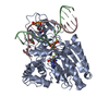


 Mass: 384.411 Da / Num. of mol.: 1 / Source method: obtained synthetically / Formula: C14H20N6O5S
 Mass: 92.094 Da / Num. of mol.: 1 / Source method: obtained synthetically / Formula: C3H8O3
 Sample preparation
 / Beamline: I02 / Wavelength: 0.97949
  Processing