 Movie
Movie Controller

[English] 日本語
 Yorodumi
- PDB-4dhy: Crystal structure of human glucokinase in complex with glucose an... -
+ Open data

- Basic information
| Entry | Database: PDB / ID: 4dhy | ||||||
|---|---|---|---|---|---|---|---|
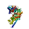
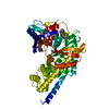
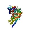
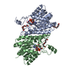
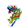
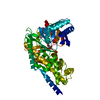
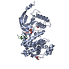
| Title | Crystal structure of human glucokinase in complex with glucose and activator | ||||||
 Components Components | Glucokinase | ||||||
 Keywords Keywords | TRANSFERASE/TRANSFERASE ACTIVATOR / DIABETES MELLITUS / DISEASE MUTATION / GLYCOLYSIS / TRANSFERASE / TRANSFERASE-TRANSFERASE ACTIVATOR complex | ||||||
| Function / homology |  Function and homology information Function and homology informationDefective GCK causes maturity-onset diabetes of the young 2 (MODY2) / glucose sensor activity / mannokinase activity / regulation of potassium ion transport / hexokinase / glucose catabolic process / fructokinase activity / glucokinase activity / glucose 6-phosphate metabolic process / NADP+ metabolic process ...Defective GCK causes maturity-onset diabetes of the young 2 (MODY2) / glucose sensor activity / mannokinase activity / regulation of potassium ion transport / hexokinase / glucose catabolic process / fructokinase activity / glucokinase activity / glucose 6-phosphate metabolic process / NADP+ metabolic process / Regulation of Glucokinase by Glucokinase Regulatory Protein / Defective TPR may confer susceptibility towards thyroid papillary carcinoma (TPC) / D-glucose binding / cellular response to leptin stimulus / calcium ion import / Glycolysis / regulation of glycolytic process / canonical glycolysis / Regulation of gene expression in beta cells / regulation of insulin secretion / negative regulation of gluconeogenesis / positive regulation of glycogen biosynthetic process / FOXO-mediated transcription of oxidative stress, metabolic and neuronal genes / response to glucose / intracellular glucose homeostasis / glycolytic process / glucose metabolic process / cellular response to insulin stimulus / positive regulation of insulin secretion / glucose homeostasis / mitochondrion / nucleoplasm / ATP binding / cytosol Similarity search - Function | ||||||
| Biological species |  Homo sapiens (human) Homo sapiens (human) | ||||||
| Method |  X-RAY DIFFRACTION / FOURIER SYNTHESIS / Resolution: 2.38 Å X-RAY DIFFRACTION / FOURIER SYNTHESIS / Resolution: 2.38 Å | ||||||
 Authors Authors | Liu, S. | ||||||
 Citation Citation | Journal: J.Biol.Chem. / Year: 2012 Title: Insights into Mechanism of Glucokinase Activation: OBSERVATION OF MULTIPLE DISTINCT PROTEIN CONFORMATIONS. Authors: Liu, S. / Ammirati, M.J. / Song, X. / Knafels, J.D. / Zhang, J. / Greasley, S.E. / Pfefferkorn, J.A. / Qiu, X. | ||||||
| History |
|
- Structure visualization
| Structure viewer | Molecule: MolmilJmol/JSmol |
|---|
- Downloads & links
-Download
| PDBx/mmCIF format | 4dhy.cif.gz | 193.1 KB | Display | PDBx/mmCIF format |
|---|---|---|---|---|
| PDB format | pdb4dhy.ent.gz | 151.3 KB | Display | PDB format |
| PDBx/mmJSON format | 4dhy.json.gz | Tree view | PDBx/mmJSON format | |
| Others |  Other downloads Other downloads |
-Validation report
| Arichive directory | https://data.pdbj.org/pub/pdb/validation_reports/dh/4dhyftp://data.pdbj.org/pub/pdb/validation_reports/dh/4dhy | HTTPS FTP |
|---|
-Related structure data
| Related structure data |  3vevC 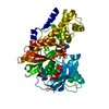 3veyC  3vf6C 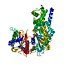 4dchC  3f9mS C: citing same article ( S: Starting model for refinement |
|---|---|
| Similar structure data |


-Links
 PDBj
PDBj


- Assembly
| Deposited unit | 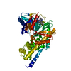
| ||||||||
|---|---|---|---|---|---|---|---|---|---|
| 1 |
| ||||||||
| Unit cell |
|
-Components
| #1: Protein | Mass: 52939.133 Da / Num. of mol.: 1 / Fragment: UNP residues 12-465 Source method: isolated from a genetically manipulated source Source: (gene. exp.) Homo sapiens (human) / Gene: GCK / Production host:  |
|---|---|
| #2: Sugar | ChemComp-GLC /   Type: D-saccharide, alpha linking / Mass: 180.156 Da / Num. of mol.: 1 Type: D-saccharide, alpha linking / Mass: 180.156 Da / Num. of mol.: 1Source method: isolated from a genetically manipulated source Formula: C6H12O6 |
| #3: Chemical | ChemComp-S41 /   Mass: 432.432 Da / Num. of mol.: 1 / Source method: obtained synthetically / Formula: C22H20N6O4 Mass: 432.432 Da / Num. of mol.: 1 / Source method: obtained synthetically / Formula: C22H20N6O4 |
| #4: Chemical | ChemComp-NA /   Mass: 22.990 Da / Num. of mol.: 1 / Source method: obtained synthetically / Formula: Na Mass: 22.990 Da / Num. of mol.: 1 / Source method: obtained synthetically / Formula: Na |
| #5: Water | ChemComp-HOH /  Mass: 18.015 Da / Num. of mol.: 181 / Source method: isolated from a natural source / Formula: H2O Mass: 18.015 Da / Num. of mol.: 181 / Source method: isolated from a natural source / Formula: H2O |
-Experimental details
-Experiment
| Experiment | Method: X-RAY DIFFRACTION / Number of used crystals: 1 |
|---|
- Sample preparation
| Crystal | Density Matthews: 2.24 Å3/Da / Density % sol: 45.12 % |
|---|---|
| Crystal grow | Temperature: 293 K / Method: vapor diffusion, hanging drop / pH: 7.5 Details: pH 7.5, VAPOR DIFFUSION, HANGING DROP, temperature 293K |
-Data collection
| Diffraction | Mean temperature: 100 K |
|---|---|
| Diffraction source | Source: ROTATING ANODE / Type: RIGAKU MICROMAX-007 / Wavelength: 1.5418 Å |
| Detector | Type: RIGAKU SATURN 944 / Detector: CCD / Date: Oct 5, 2010 |
| Radiation | Protocol: SINGLE WAVELENGTH / Monochromatic (M) / Laue (L): M / Scattering type: x-ray |
| Radiation wavelength | Wavelength: 1.5418 Å / Relative weight: 1 |
| Reflection | Resolution: 2.38→52.9 Å / Num. obs: 18951 / % possible obs: 96.8 % / Observed criterion σ(I): 2 / Redundancy: 5.3 % / Biso Wilson estimate: 35.87 Å2 / Rmerge(I) obs: 0.109 / Net I/σ(I): 12.5 |
| Reflection shell | Resolution: 2.38→2.51 Å / Redundancy: 3.8 % / Rmerge(I) obs: 0.403 / Mean I/σ(I) obs: 3.1 / Num. unique all: 2562 / % possible all: 92.5 |
- Processing
| Software | Name: BUSTER / Version: 2.11.2 / Classification: refinement | ||||||||||||||||||||||||||||||||||||||||||||||||||||||||||||||||||||||||||||||||||||||||||||||||||||||||||||||||||
|---|---|---|---|---|---|---|---|---|---|---|---|---|---|---|---|---|---|---|---|---|---|---|---|---|---|---|---|---|---|---|---|---|---|---|---|---|---|---|---|---|---|---|---|---|---|---|---|---|---|---|---|---|---|---|---|---|---|---|---|---|---|---|---|---|---|---|---|---|---|---|---|---|---|---|---|---|---|---|---|---|---|---|---|---|---|---|---|---|---|---|---|---|---|---|---|---|---|---|---|---|---|---|---|---|---|---|---|---|---|---|---|---|---|---|---|
| Refinement | Method to determine structure: FOURIER SYNTHESIS Starting model: 3F9M Resolution: 2.38→52.9 Å / Cor.coef. Fo:Fc: 0.9292 / Cor.coef. Fo:Fc free: 0.8868 / SU R Cruickshank DPI: 0.435 / Cross valid method: THROUGHOUT / σ(F): 0 / Stereochemistry target values: Engh & Huber
| ||||||||||||||||||||||||||||||||||||||||||||||||||||||||||||||||||||||||||||||||||||||||||||||||||||||||||||||||||
| Displacement parameters | Biso mean: 29.19 Å2
| ||||||||||||||||||||||||||||||||||||||||||||||||||||||||||||||||||||||||||||||||||||||||||||||||||||||||||||||||||
| Refine analyze | Luzzati coordinate error obs: 0.288 Å | ||||||||||||||||||||||||||||||||||||||||||||||||||||||||||||||||||||||||||||||||||||||||||||||||||||||||||||||||||
| Refinement step | Cycle: LAST / Resolution: 2.38→52.9 Å
| ||||||||||||||||||||||||||||||||||||||||||||||||||||||||||||||||||||||||||||||||||||||||||||||||||||||||||||||||||
| Refine LS restraints |
| ||||||||||||||||||||||||||||||||||||||||||||||||||||||||||||||||||||||||||||||||||||||||||||||||||||||||||||||||||
| LS refinement shell | Resolution: 2.38→2.51 Å / Total num. of bins used: 10
| ||||||||||||||||||||||||||||||||||||||||||||||||||||||||||||||||||||||||||||||||||||||||||||||||||||||||||||||||||
| Refinement TLS params. | Method: refined / Origin x: -27.5812 Å / Origin y: -0.8077 Å / Origin z: 9.5699 Å
|
