 Movie
Movie Controller

[English] 日本語
 Yorodumi
- PDB-4dgb: TRIMCyp cyclophilin domain from Macaca mulatta: HIV-2 CA cyclophi... -
+ Open data

- Basic information
| Entry | Database: PDB / ID: 4dgb | ||||||
|---|---|---|---|---|---|---|---|
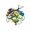
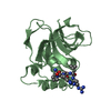
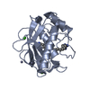
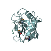
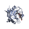
| Title | TRIMCyp cyclophilin domain from Macaca mulatta: HIV-2 CA cyclophilin-binding loop complex | ||||||
 Components Components |
| ||||||
 Keywords Keywords | ISOMERASE/VIRAL PROTEIN / anti-viral protein / ISOMERASE-VIRAL PROTEIN complex | ||||||
| Function / homology |  Function and homology information Function and homology informationnegative regulation of protein K48-linked ubiquitination / regulation of apoptotic signaling pathway / cell adhesion molecule production / lipid droplet organization / negative regulation of viral life cycle / heparan sulfate binding / regulation of viral genome replication / leukocyte chemotaxis / negative regulation of stress-activated MAPK cascade / activation of protein kinase B activity ...negative regulation of protein K48-linked ubiquitination / regulation of apoptotic signaling pathway / cell adhesion molecule production / lipid droplet organization / negative regulation of viral life cycle / heparan sulfate binding / regulation of viral genome replication / leukocyte chemotaxis / negative regulation of stress-activated MAPK cascade / activation of protein kinase B activity / endothelial cell activation / protein peptidyl-prolyl isomerization / cyclosporin A binding / viral budding via host ESCRT complex / negative regulation of protein phosphorylation / negative regulation of protein kinase activity / negative regulation of oxidative stress-induced intrinsic apoptotic signaling pathway / positive regulation of viral genome replication / neutrophil chemotaxis / : / peptidylprolyl isomerase / positive regulation of protein secretion / peptidyl-prolyl cis-trans isomerase activity / platelet activation / positive regulation of protein phosphorylation / RING-type E3 ubiquitin transferase / platelet aggregation / integrin binding / host multivesicular body / autophagy / ubiquitin protein ligase activity / protein folding / viral nucleocapsid / cellular response to oxidative stress / defense response to virus / positive regulation of MAPK cascade / protein ubiquitination / viral translational frameshifting / innate immune response / apoptotic process / host cell plasma membrane / host cell nucleus / virion membrane / structural molecule activity / protein-containing complex / RNA binding / extracellular region / zinc ion binding / nucleus / cytoplasm / cytosol Similarity search - Function | ||||||
| Biological species |   Human immunodeficiency virus 2 Human immunodeficiency virus 2 | ||||||
| Method |  X-RAY DIFFRACTION / MOLECULAR REPLACEMENT / Resolution: 1.702 Å X-RAY DIFFRACTION / MOLECULAR REPLACEMENT / Resolution: 1.702 Å | ||||||
 Authors Authors | Caines, M.E.C. / Bichel, K. / Price, A.J. / McEwan, W.A. / James, L.C. | ||||||
 Citation Citation | Journal: Nat.Struct.Mol.Biol. / Year: 2012 Title: Diverse HIV viruses are targeted by a conformationally dynamic antiviral. Authors: Caines, M.E. / Bichel, K. / Price, A.J. / McEwan, W.A. / Towers, G.J. / Willett, B.J. / Freund, S.M. / James, L.C. | ||||||
| History |
|
- Structure visualization
| Structure viewer | Molecule: MolmilJmol/JSmol |
|---|
- Downloads & links
-Download
| PDBx/mmCIF format | 4dgb.cif.gz | 81 KB | Display | PDBx/mmCIF format |
|---|---|---|---|---|
| PDB format | pdb4dgb.ent.gz | 60 KB | Display | PDB format |
| PDBx/mmJSON format | 4dgb.json.gz | Tree view | PDBx/mmJSON format | |
| Others |  Other downloads Other downloads |
-Validation report
| Arichive directory | https://data.pdbj.org/pub/pdb/validation_reports/dg/4dgbftp://data.pdbj.org/pub/pdb/validation_reports/dg/4dgb | HTTPS FTP |
|---|
-Related structure data
| Related structure data |  4dgaC  4dgcC 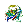 4dgdC  4dgeC  2wlwS C: citing same article ( S: Starting model for refinement |
|---|---|
| Similar structure data |


-Links
 PDBj
PDBj


- Assembly
| Deposited unit | 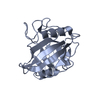
| ||||||||
|---|---|---|---|---|---|---|---|---|---|
| 1 |
| ||||||||
| Unit cell |
|
-Components
| #1: Protein | Mass: 18016.473 Da / Num. of mol.: 1 / Fragment: cyclophilin domain (UNP residues 304-468) Source method: isolated from a genetically manipulated source Source: (gene. exp.)  References: UniProt: B0LJC8, UniProt: P62940*PLUS, peptidylprolyl isomerase |
|---|---|
| #2: Protein/peptide | Mass: 550.647 Da / Num. of mol.: 1 / Fragment: cyclophilin-binding loop (UNP residues 221-226) / Source method: obtained synthetically / Source: (synth.) Human immunodeficiency virus 2 / References: UniProt: P04590 |
| #3: Water | ChemComp-HOH /  Mass: 18.015 Da / Num. of mol.: 225 / Source method: isolated from a natural source / Formula: H2O Mass: 18.015 Da / Num. of mol.: 225 / Source method: isolated from a natural source / Formula: H2O |
-Experimental details
-Experiment
| Experiment | Method: X-RAY DIFFRACTION / Number of used crystals: 1 |
|---|
- Sample preparation
| Crystal | Density Matthews: 1.92 Å3/Da / Density % sol: 36.02 % |
|---|---|
| Crystal grow | Temperature: 290 K / Method: vapor diffusion, sitting drop / pH: 8.3 Details: 2.35 M ammonium phosphate dibasic, 0.1 M Tris-HCl, pH 8.3, VAPOR DIFFUSION, SITTING DROP, temperature 290K |
-Data collection
| Diffraction | Mean temperature: 100 K | ||||||||||||||||||||||||||||||||||||||||||||||||||||||||||||||||||||||||||||||||||||||||
|---|---|---|---|---|---|---|---|---|---|---|---|---|---|---|---|---|---|---|---|---|---|---|---|---|---|---|---|---|---|---|---|---|---|---|---|---|---|---|---|---|---|---|---|---|---|---|---|---|---|---|---|---|---|---|---|---|---|---|---|---|---|---|---|---|---|---|---|---|---|---|---|---|---|---|---|---|---|---|---|---|---|---|---|---|---|---|---|---|---|
| Diffraction source | Source: ROTATING ANODE / Type: RIGAKU RU300 / Wavelength: 1.5418 Å | ||||||||||||||||||||||||||||||||||||||||||||||||||||||||||||||||||||||||||||||||||||||||
| Detector | Type: MAR scanner 345 mm plate / Detector: IMAGE PLATE / Date: Jul 9, 2009 | ||||||||||||||||||||||||||||||||||||||||||||||||||||||||||||||||||||||||||||||||||||||||
| Radiation | Protocol: SINGLE WAVELENGTH / Monochromatic (M) / Laue (L): M / Scattering type: x-ray | ||||||||||||||||||||||||||||||||||||||||||||||||||||||||||||||||||||||||||||||||||||||||
| Radiation wavelength | Wavelength: 1.5418 Å / Relative weight: 1 | ||||||||||||||||||||||||||||||||||||||||||||||||||||||||||||||||||||||||||||||||||||||||
| Reflection | Resolution: 1.702→71.08 Å / Num. all: 16284 / Num. obs: 15839 / % possible obs: 95 % / Redundancy: 2.3 % / Biso Wilson estimate: 17.04 Å2 / Rsym value: 0.037 / Net I/σ(I): 14.9 | ||||||||||||||||||||||||||||||||||||||||||||||||||||||||||||||||||||||||||||||||||||||||
| Reflection shell | Diffraction-ID: 1
|
- Processing
| Software |
| ||||||||||||||||||||||||||||||||||||||||||||||||||||||||||||
|---|---|---|---|---|---|---|---|---|---|---|---|---|---|---|---|---|---|---|---|---|---|---|---|---|---|---|---|---|---|---|---|---|---|---|---|---|---|---|---|---|---|---|---|---|---|---|---|---|---|---|---|---|---|---|---|---|---|---|---|---|---|
| Refinement | Method to determine structure: MOLECULAR REPLACEMENT Starting model: PDB ENTRY 2WLW Resolution: 1.702→30.34 Å / Cor.coef. Fo:Fc: 0.958 / Cor.coef. Fo:Fc free: 0.943 / WRfactor Rfree: 0.2115 / WRfactor Rwork: 0.1775 / Occupancy max: 1 / Occupancy min: 0.5 / FOM work R set: 0.8878 / SU B: 3.89 / SU ML: 0.068 / SU R Cruickshank DPI: 0.128 / SU Rfree: 0.117 / Cross valid method: THROUGHOUT / σ(F): 0 / ESU R: 0.128 / ESU R Free: 0.117 / Stereochemistry target values: MAXIMUM LIKELIHOOD Details: HYDROGENS HAVE BEEN ADDED IN THE RIDING POSITIONS U VALUES : WITH TLS ADDED
| ||||||||||||||||||||||||||||||||||||||||||||||||||||||||||||
| Solvent computation | Ion probe radii: 0.8 Å / Shrinkage radii: 0.8 Å / VDW probe radii: 1.2 Å / Solvent model: MASK | ||||||||||||||||||||||||||||||||||||||||||||||||||||||||||||
| Displacement parameters | Biso max: 56.33 Å2 / Biso mean: 18.2559 Å2 / Biso min: 9.58 Å2
| ||||||||||||||||||||||||||||||||||||||||||||||||||||||||||||
| Refinement step | Cycle: LAST / Resolution: 1.702→30.34 Å
| ||||||||||||||||||||||||||||||||||||||||||||||||||||||||||||
| Refine LS restraints |
| ||||||||||||||||||||||||||||||||||||||||||||||||||||||||||||
| LS refinement shell | Resolution: 1.702→1.746 Å / Total num. of bins used: 20
| ||||||||||||||||||||||||||||||||||||||||||||||||||||||||||||
| Refinement TLS params. | Method: refined / Origin x: 9.3657 Å / Origin y: -0.2112 Å / Origin z: 7.0043 Å
|
