- PDB-4dck: Crystal structure of the C-terminus of voltage-gated sodium chann... -
+
Open data
ID or keywords:
Loading...
-
Basic information
Entry
Database: PDB / ID: 4dck
Title
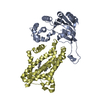
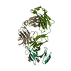
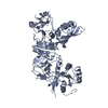
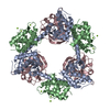
Crystal structure of the C-terminus of voltage-gated sodium channel in complex with FGF13 and CaM
Components
Calmodulin
Fibroblast growth factor 13
Sodium channel protein type 5 subunit alpha
Keywords
TRANSPORT PROTEIN/SIGNALING PROTEIN / IQ-motif / EF-hand / Voltage-gated Sodium Channel regulation / Nav1.5 CTD binds to FGF13 and CaM. CaM binds to Ca2+. / TRANSPORT PROTEIN-TRANSPORT PROTEIN REGULATOR-SIGNALING PROTEIN complex / TRANSPORT PROTEIN-SIGNALING PROTEIN complex
Function / homology
Function and homology information
establishment of neuroblast polarity / voltage-gated sodium channel activity involved in AV node cell action potential / voltage-gated sodium channel activity involved in bundle of His cell action potential / voltage-gated sodium channel activity involved in Purkinje myocyte action potential / voltage-gated sodium channel activity involved in SA node cell action potential / regulation of cardiac muscle cell action potential involved in regulation of contraction / bundle of His cell action potential / regulation of ventricular cardiac muscle cell membrane depolarization / AV node cell action potential / SA node cell action potential ...establishment of neuroblast polarity / voltage-gated sodium channel activity involved in AV node cell action potential / voltage-gated sodium channel activity involved in bundle of His cell action potential / voltage-gated sodium channel activity involved in Purkinje myocyte action potential / voltage-gated sodium channel activity involved in SA node cell action potential / regulation of cardiac muscle cell action potential involved in regulation of contraction / bundle of His cell action potential / regulation of ventricular cardiac muscle cell membrane depolarization / AV node cell action potential / SA node cell action potential / AV node cell to bundle of His cell communication / membrane depolarization during SA node cell action potential / negative regulation of collateral sprouting / positive regulation of voltage-gated sodium channel activity / response to denervation involved in regulation of muscle adaptation / membrane depolarization during atrial cardiac muscle cell action potential / voltage-gated sodium channel activity involved in cardiac muscle cell action potential / regulation of atrial cardiac muscle cell membrane repolarization / cardiac ventricle development / brainstem development / : / : / : / : / regulation of atrial cardiac muscle cell membrane depolarization / positive regulation of action potential / atrial cardiac muscle cell action potential / positive regulation of protein autophosphorylation / : / cardiac conduction system development / membrane depolarization during AV node cell action potential / membrane depolarization during bundle of His cell action potential / telencephalon development / membrane depolarization during cardiac muscle cell action potential / negative regulation of peptidyl-threonine phosphorylation / membrane depolarization during Purkinje myocyte cell action potential / positive regulation of sodium ion transport / membrane depolarization during action potential / negative regulation of microtubule depolymerization / regulation of sodium ion transmembrane transport / : / ventricular cardiac muscle cell action potential / type 3 metabotropic glutamate receptor binding / regulation of ventricular cardiac muscle cell membrane repolarization / cardiac muscle cell action potential involved in contraction / voltage-gated sodium channel complex / regulation of cardiac muscle cell contraction / inhibitory synapse assembly / positive regulation of peptidyl-threonine phosphorylation / positive regulation of DNA binding / CaM pathway / Cam-PDE 1 activation / voltage-gated sodium channel activity / Sodium/Calcium exchangers / Interaction between L1 and Ankyrins / ankyrin binding / Calmodulin induced events / Reduction of cytosolic Ca++ levels / Activation of Ca-permeable Kainate Receptor / CREB1 phosphorylation through the activation of CaMKII/CaMKK/CaMKIV cascasde / Loss of phosphorylation of MECP2 at T308 / CREB1 phosphorylation through the activation of Adenylate Cyclase / negative regulation of high voltage-gated calcium channel activity / PKA activation / CaMK IV-mediated phosphorylation of CREB / Glycogen breakdown (glycogenolysis) / response to corticosterone / negative regulation of ryanodine-sensitive calcium-release channel activity / Activation of RAC1 downstream of NMDARs / organelle localization by membrane tethering / CLEC7A (Dectin-1) induces NFAT activation / odontogenesis of dentin-containing tooth / : / autophagosome membrane docking / regulation of synaptic vesicle exocytosis / negative regulation of calcium ion export across plasma membrane / regulation of ryanodine-sensitive calcium-release channel activity / regulation of cardiac muscle cell action potential / presynaptic endocytosis / cerebral cortex cell migration / Synthesis of IP3 and IP4 in the cytosol / sodium ion transport / positive regulation of protein serine/threonine kinase activity / Phase 0 - rapid depolarisation / Negative regulation of NMDA receptor-mediated neuronal transmission / microtubule polymerization / Unblocking of NMDA receptors, glutamate binding and activation / calcineurin-mediated signaling / RHO GTPases activate PAKs / nitric-oxide synthase binding / regulation of heart rate by cardiac conduction / regulation of cell communication by electrical coupling involved in cardiac conduction / fibroblast growth factor binding / Ion transport by P-type ATPases / adenylate cyclase binding / Uptake and function of anthrax toxins / protein phosphatase activator activity / intercalated disc / Long-term potentiation / lateral plasma membrane Similarity search - Function
iswi atpase / Voltage gated sodium channel, alpha-5 subunit / Voltage-gated Na+ ion channel, cytoplasmic domain / Cytoplasmic domain of voltage-gated Na+ ion channel / HBGF/FGF family signature. / Fibroblast growth factor family / Fibroblast growth factor / Acidic and basic fibroblast growth factor family. / Sodium ion transport-associated / Voltage-gated sodium channel alpha subunit, inactivation gate ...iswi atpase / Voltage gated sodium channel, alpha-5 subunit / Voltage-gated Na+ ion channel, cytoplasmic domain / Cytoplasmic domain of voltage-gated Na+ ion channel / HBGF/FGF family signature. / Fibroblast growth factor family / Fibroblast growth factor / Acidic and basic fibroblast growth factor family. / Sodium ion transport-associated / Voltage-gated sodium channel alpha subunit, inactivation gate / : / Sodium ion transport-associated / SCN5A-like, C-terminal IQ motif / Voltage gated sodium channel, alpha subunit / Cytokine IL1/FGF / Voltage-gated cation channel calcium and sodium / Trefoil (Acidic Fibroblast Growth Factor, subunit A) - #50 / Trefoil (Acidic Fibroblast Growth Factor, subunit A) / Trefoil / Voltage-dependent channel domain superfamily / EF-hand / Recoverin; domain 1 / : / Single alpha-helices involved in coiled-coils or other helix-helix interfaces / EF-hand domain pair / EF-hand, calcium binding motif / EF-Hand 1, calcium-binding site / EF-hand calcium-binding domain. / EF-hand calcium-binding domain profile. / EF-hand domain / EF-hand domain pair / Ion transport domain / Ion transport protein / Up-down Bundle / Orthogonal Bundle / Mainly Beta / Mainly Alpha Similarity search - Domain/homology
Calmodulin-1 / Calmodulin-3 / Sodium channel protein type 5 subunit alpha / Fibroblast growth factor 13 Similarity search - Component
Biological species
Homo sapiens (human)
Method
X-RAY DIFFRACTION / SYNCHROTRON / SAD / Resolution: 2.2 Å
In the structure databanks used in Yorodumi, some data are registered as the other names, "COVID-19 virus" and "2019-nCoV". Here are the details of the virus and the list of structure data.
Jan 31, 2019. EMDB accession codes are about to change! (news from PDBe EMDB page)
EMDB accession codes are about to change! (news from PDBe EMDB page)
The allocation of 4 digits for EMDB accession codes will soon come to an end. Whilst these codes will remain in use, new EMDB accession codes will include an additional digit and will expand incrementally as the available range of codes is exhausted. The current 4-digit format prefixed with “EMD-” (i.e. EMD-XXXX) will advance to a 5-digit format (i.e. EMD-XXXXX), and so on. It is currently estimated that the 4-digit codes will be depleted around Spring 2019, at which point the 5-digit format will come into force.
The EM Navigator/Yorodumi systems omit the EMD- prefix.
Related info.:Q: What is EMD? / ID/Accession-code notation in Yorodumi/EM Navigator
Yorodumi is a browser for structure data from EMDB, PDB, SASBDB, etc.
This page is also the successor to EM Navigator detail page, and also detail information page/front-end page for Omokage search.
The word "yorodu" (or yorozu) is an old Japanese word meaning "ten thousand". "mi" (miru) is to see.
Related info.:EMDB / PDB / SASBDB / Comparison of 3 databanks / Yorodumi Search / Aug 31, 2016. New EM Navigator & Yorodumi / Yorodumi Papers / Jmol/JSmol / Function and homology information / Changes in new EM Navigator and Yorodumi
 Movie
Movie Controller

 Yorodumi
 Open data
 Basic information
 Components
 Keywords
 Function and homology information
 Homo sapiens (human)
 X-RAY DIFFRACTION /
  Authors
 Citation
 Structure visualization
 Downloads & links
 Other downloads
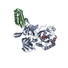


 PDBj
PDBj


 Assembly


 Mass: 24.305 Da / Num. of mol.: 3 / Source method: obtained synthetically / Formula: Mg
 Mass: 18.015 Da / Num. of mol.: 202 / Source method: isolated from a natural source / Formula: H2O
 Sample preparation
 Processing