- PDB-4d6y: Crystal structure of the receiver domain of NtrX from Brucella ab... -
+
Open data
ID or keywords:
Loading...
-
Basic information
Entry
Database: PDB / ID: 4d6y
Title
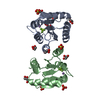
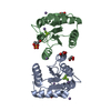
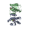
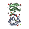
Crystal structure of the receiver domain of NtrX from Brucella abortus in complex with beryllofluoride and magnesium
Components
BACTERIAL REGULATORY, FIS FAMILY PROTEIN
Keywords
SIGNALING PROTEIN / BRUCELLOSIS / TWO-COMPONENT SYSTEM / RESPONSE REGULATOR / REC DOMAIN / MICROAEROBISIS
Function / homology
Function and homology information
phosphorelay signal transduction system / sequence-specific DNA binding / regulation of DNA-templated transcription / DNA-templated transcription / ATP binding / metal ion binding Similarity search - Function
Sigma-54 interaction domain, conserved site / Sigma-54 interaction domain C-terminal part signature. / Sigma-54 interaction domain, ATP-binding site 2 / Sigma-54 interaction domain ATP-binding region B signature. / : / AAA+ ATPase lid domain / Sigma-54 interaction domain profile. / Sigma-54 interaction domain / RNA polymerase sigma factor 54 interaction domain / DNA binding HTH domain, Fis-type ...Sigma-54 interaction domain, conserved site / Sigma-54 interaction domain C-terminal part signature. / Sigma-54 interaction domain, ATP-binding site 2 / Sigma-54 interaction domain ATP-binding region B signature. / : / AAA+ ATPase lid domain / Sigma-54 interaction domain profile. / Sigma-54 interaction domain / RNA polymerase sigma factor 54 interaction domain / DNA binding HTH domain, Fis-type / Bacterial regulatory protein, Fis family / Response regulator receiver domain / cheY-homologous receiver domain / Signal transduction response regulator, receiver domain / Response regulatory domain profile. / CheY-like superfamily / Response regulator / Homeobox-like domain superfamily / ATPases associated with a variety of cellular activities / AAA+ ATPase domain / Rossmann fold / P-loop containing nucleoside triphosphate hydrolase / 3-Layer(aba) Sandwich / Alpha Beta Similarity search - Domain/homology
In the structure databanks used in Yorodumi, some data are registered as the other names, "COVID-19 virus" and "2019-nCoV". Here are the details of the virus and the list of structure data.
Jan 31, 2019. EMDB accession codes are about to change! (news from PDBe EMDB page)
EMDB accession codes are about to change! (news from PDBe EMDB page)
The allocation of 4 digits for EMDB accession codes will soon come to an end. Whilst these codes will remain in use, new EMDB accession codes will include an additional digit and will expand incrementally as the available range of codes is exhausted. The current 4-digit format prefixed with “EMD-” (i.e. EMD-XXXX) will advance to a 5-digit format (i.e. EMD-XXXXX), and so on. It is currently estimated that the 4-digit codes will be depleted around Spring 2019, at which point the 5-digit format will come into force.
The EM Navigator/Yorodumi systems omit the EMD- prefix.
Related info.:Q: What is EMD? / ID/Accession-code notation in Yorodumi/EM Navigator
Yorodumi is a browser for structure data from EMDB, PDB, SASBDB, etc.
This page is also the successor to EM Navigator detail page, and also detail information page/front-end page for Omokage search.
The word "yorodu" (or yorozu) is an old Japanese word meaning "ten thousand". "mi" (miru) is to see.
Related info.:EMDB / PDB / SASBDB / Comparison of 3 databanks / Yorodumi Search / Aug 31, 2016. New EM Navigator & Yorodumi / Yorodumi Papers / Jmol/JSmol / Function and homology information / Changes in new EM Navigator and Yorodumi
 Movie
Movie Controller

 Yorodumi
 Open data
 Basic information
 Components
 Keywords
 Function and homology information
 BRUCELLA ABORTUS (bacteria)
 X-RAY DIFFRACTION /
  Authors
 Citation
 Structure visualization
 Downloads & links
 Other downloads
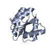


 PDBj
PDBj


 Assembly

 Mass: 66.007 Da / Num. of mol.: 2 / Source method: obtained synthetically / Formula: BeF3
 Mass: 24.305 Da / Num. of mol.: 2 / Source method: obtained synthetically / Formula: Mg
 Mass: 18.015 Da / Num. of mol.: 106 / Source method: isolated from a natural source / Formula: H2O
 Sample preparation
 / Beamline: PROXIMA 1 / Wavelength: 0.97625
  Processing