Monochromator: MIRROR / Protocol: SINGLE WAVELENGTH / Monochromatic (M) / Laue (L): M / Scattering type: x-ray
Radiation wavelength
Wavelength: 1.5418 Å / Relative weight: 1
Reflection
Resolution: 1.7→50 Å / Num. obs: 21903 / % possible obs: 95.7 % / Observed criterion σ(I): 0 / Redundancy: 8.7 % / Biso Wilson estimate: 13 Å2 / Rmerge(I) obs: 0.06 / Net I/σ(I): 29.7
Reflection shell
Resolution: 1.7→1.75 Å / Redundancy: 3.3 % / Rmerge(I) obs: 0.81 / Mean I/σ(I) obs: 2 / % possible all: 71.1
-
Processing
Software
Name
Version
Classification
REFMAC
5.8.0073
refinement
XDS
datareduction
XDS
datascaling
PHASER
phasing
Refinement
Method to determine structure: MOLECULAR REPLACEMENT / Resolution: 1.7→53.03 Å / Cor.coef. Fo:Fc: 0.957 / Cor.coef. Fo:Fc free: 0.954 / SU B: 6.154 / SU ML: 0.087 / Cross valid method: THROUGHOUT / ESU R: 0.109 / ESU R Free: 0.101 / Stereochemistry target values: MAXIMUM LIKELIHOOD Details: HYDROGENS HAVE BEEN ADDED IN THE RIDING POSITIONS. U VALUES WITH TLS ADDED.
Rfactor
Num. reflection
% reflection
Selection details
Rfree
0.19722
1094
5 %
RANDOM
Rwork
0.17364
-
-
-
obs
0.17486
20809
95.71 %
-
Solvent computation
Ion probe radii: 0.8 Å / Shrinkage radii: 0.8 Å / VDW probe radii: 1.2 Å / Solvent model: BABINET MODEL WITH MASK
 Movie
Movie Controller


 Open data
 Basic information
 Components
 Keywords
 Function and homology information
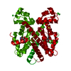
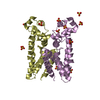
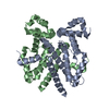
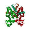
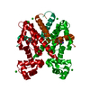
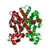
 PROTEUS MIRABILIS (bacteria)
 X-RAY DIFFRACTION /
  Authors
 Citation
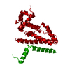
 Structure visualization
 Downloads & links
 Other downloads
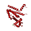


 PDBj
PDBj Assembly
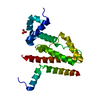

 Mass: 96.063 Da / Num. of mol.: 1 / Source method: obtained synthetically / Formula: SO4
 Mass: 18.015 Da / Num. of mol.: 192 / Source method: isolated from a natural source / Formula: H2O
 Sample preparation
 Processing